
Page 43 - Read Online
P. 43
Page 8 of 14 Grewal et al. Plast Aesthet Res 2021;8:37 https://dx.doi.org/10.20517/2347-9264.2021.43
weeks. Eight weeks after their operation they can resume a regular diet. They will follow up with their
prosthodontist for frequent outpatient visits and prosthesis adjustments/maintenance and receive their final
prosthesis at 4 months.
CASE REPORT
We performed the procedure as described above in August 2019 [Table 1]. Our index patient was a 51-year-
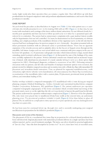
old male who initially presented in 2011 to the treating OMS with an OKC [Figure 4]. At that time, he was
treated with enucleation and curettage of the lesion without dental extractions. He was followed closely for 7
months post-operatively and then was lost to follow up until early 2019 when he re-presented upon self-
referral for re-evaluation. The patient had no symptomatic complaints with a past medical history notable
only for hypertension that was well controlled. On exam, he demonstrated no facial asymmetry, no obvious
swelling, and a slight paresthesia of the mandibular division of the trigeminal nerve on the left side that he
reported had been unchanged since his previous surgery in 2011. Intraorally - the patient presented with an
intact permanent dentition with no advanced caries or periodontal disease. There was no apparent
expansion of the alveolar process and no palpable defect in the buccal or lingual cortex throughout the
mandible. Soft tissue changes and scar were noted which were consistent with prior surgery in the area of
his lower left quadrant. A 2-D panoramic radiograph was taken which demonstrated a large, mesial-distal
extending, multi-loculated, radiolucent lesion [Figure 5]. At that point the sum of the findings of the exam
were carefully explained to the patient. Written informed consent was obtained and an incisional biopsy
was obtained, with simultaneous placement of a nasal cannula tubing to serve as a drain (given high
suspicion for OKC). Histological diagnosis confirmed a recurrence of an OKC. Following extensive
discussion with the patient, and all risks and benefits of multiple treatment options were reviewed, the
patient elected for definitive surgical resection and reconstruction with a fibula free flap with immediate DI
placement and immediate dental restoration. Thus, we planned for segmental mandibulectomy, tooth
extractions, right inferior alveolar nerve lateralization, left osteocutaneous free fibular flap reconstruction,
reconstruction of the mandibular defect with a custom plate, DI placement, provisional dental prosthesis
delivery, and immediate loading of DIs.
Further workup included a computed tomography (CT) maxillofacial with 0.5 mm slicing per surgical
planning company (Medical Modeling Inc., Golden, Colorado) and customized medical device company
(Stryker Corporation, Kalamazoo, MI) guidelines [Figure 6]. The patient underwent pre-operative
computed tomography angiography of the lower extremities which revealed distal narrowing of the
peroneal vessels, more so on the right than the left, so it was decided to harvest the graft from his left side.
The patient underwent his operation without major complication [Figure 7]. However of the five planned
DIs, only three were placed due to insufficient fibula bone width at the selected sites, an unforeseen
planning error. The provisional prosthesis included 10 teeth [Figure 8]. Other than failure to place all
planned DIs, he suffered no complications intra-operatively or post-operatively and was discharged seven
days later on a puree diet.
He has been seen for continual follow up, through 2021 and is currently undergoing post-surgical
modifications in preparation for his definitive prosthesis [Figure 9].
Discussion and review of the literature
The placement of DIs in a vascularized free tissue flap in preparation for a delayed dental prosthesis has
been well described. Implant placement with immediate prosthesis delivery in a single operation has been
seldom reported, and is a novel treatment paradigm. The index case above details the practical application
of “Jaw in a Day.” Since the first description by Levine et al. in 2013, there have been 20 recorded cases,
[1]