
Page 75 - Read Online
P. 75
Neligan. Plast Aesthet Res 2021;8:45 https://dx.doi.org/10.20517/2347-9264.2021.39 Page 7 of 9
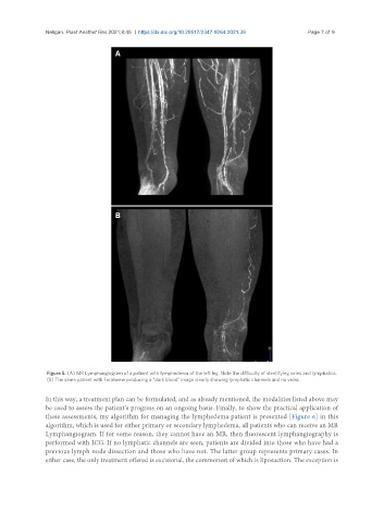
Figure 5. (A) MR Lymphangiogram of a patient with lymphedema of the left leg. Note the difficulty of identifying veins and lymphatics.
(B) The same patient with Feraheme producing a “dark blood” image clearly showing lymphatic channels and no veins.
In this way, a treatment plan can be formulated, and as already mentioned, the modalities listed above may
be used to assess the patient’s progress on an ongoing basis. Finally, to show the practical application of
these assessments, my algorithm for managing the lymphedema patient is presented [Figure 6] in this
algorithm, which is used for either primary or secondary lymphedema, all patients who can receive an MR
Lymphangiogram. If for some reason, they cannot have an MR, then fluorescent lymphangiography is
performed with ICG. If no lymphatic channels are seen, patients are divided into those who have had a
previous lymph node dissection and those who have not. The latter group represents primary cases. In
either case, the only treatment offered is excisional, the commonest of which is liposuction. The exception is