
Page 72 - Read Online
P. 72
Page 4 of 9 Neligan. Plast Aesthet Res 2021;8:45 https://dx.doi.org/10.20517/2347-9264.2021.39
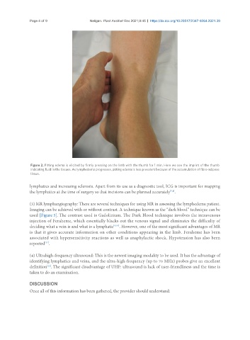
Figure 2. Pitting edema is elicited by firmly pressing on the limb with the thumb for 1 min. Here we see the imprint of the thumb
indicating fluid in the tissues. As lymphedema progresses, pitting edema is less prevalent because of the accumulation of fibro-adipose
tissue.
lymphatics and increasing sclerosis. Apart from its use as a diagnostic tool, ICG is important for mapping
[7,8]
the lymphatics at the time of surgery so that incisions can be planned accurately .
(3) MR lymphangiography: There are several techniques for using MR in assessing the lymphedema patient.
Imaging can be achieved with or without contrast. A technique known as the “dark blood” technique can be
used [Figure 5]. The contrast used is Gadolinium. The Dark Blood technique involves the intravenous
injection of Feraheme, which essentially blacks out the venous signal and eliminates the difficulty of
deciding what a vein is and what is a lymphatic [9,10] . However, one of the most significant advantages of MR
is that it gives accurate information on other conditions appearing in the limb. Feraheme has been
associated with hypersensitivity reactions as well as anaphylactic shock. Hypotension has also been
reported .
[11]
(4) Ultrahigh-frequency ultrasound: This is the newest imaging modality to be used. It has the advantage of
identifying lymphatics and veins, and the ultra-high-frequency (up to 70 MHz) probes give an excellent
definition . The significant disadvantage of UHF: ultrasound is lack of user-friendliness and the time is
[12]
taken to do an examination.
DISCUSSION
Once all of this information has been gathered, the provider should understand: