
Page 71 - Read Online
P. 71
Neligan. Plast Aesthet Res 2021;8:45 https://dx.doi.org/10.20517/2347-9264.2021.39 Page 3 of 9
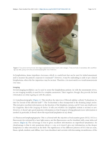
Figure 1. This patient with extreme late-stage lymphedema shows trophic skin changes. There are folds of edematous skin and thick
rigorous skin, giving rise to fissures and increasing the risk of infection.
In lymphedema, tissue impedance decreases, which is a useful test that can be used for initial assessment
[5]
and to measure the patient’s response to treatment . However, it may be misleading in early or pre-clinical
lymphedema, where the bio-impedence may be normal. Therefore, it is most useful as a trend measurement
over time.
Imaging
Several imaging modalities can be used to assess the lymphedema patient. As with the assessments above,
no one imaging modality is used for the complete assessment. Taken together, though, they provide the best
information on what is going on with the patient.
(1) Lymphoscintigraphy [Figure 3]: This involves the injection of filtered sulphur colloid, Technetium 99,
into the dermis of the affected limb . The Technetium is then transported to the draining lymph nodes.
[6,7]
This text gives excellent information on the function of the lymphatic system, and if I have any doubt as to
the diagnosis, this is the imaging of choice. It tells you whether the lymphatic system is normal or not.
However, it doesn’t give good anatomic information so that if surgery is being planned, more information is
needed. In particular, it doesn’t show the superficial lymphatics in any detail.
(2) Fluorescent lymphangiography: This is achieved with the injection of indocyanine green (ICG). ICG is a
fluorescent dye activated by a laser light source, and the fluorescence can be visualized with a near infra-red
camera [Figure 4]. The advantage is that it gives excellent information on superficial lymphatics. Its
disadvantage is that it only penetrates about 1.5 cm, so it doesn’t give any information on the deeper
lymphatics or other structures in the limb. The significance of the different patterns of ICG that are seen,
linear, splash, stardust, and diffuse, have been elucidated and correlate with decreasing circumference of the