
Page 28 - Read Online
P. 28
Chen et al. Plast Aesthet Res 2021;8:36 https://dx.doi.org/10.20517/2347-9264.2021.33 Page 3 of 6
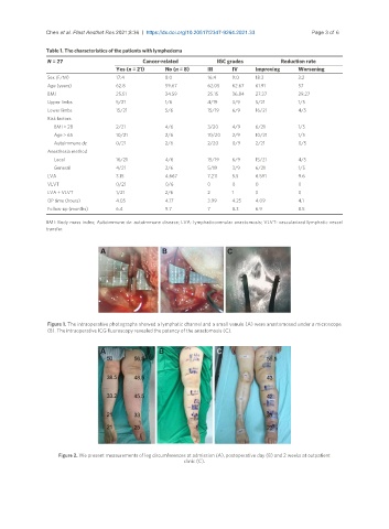
Table 1. The characteristics of the patients with lymphedema
N = 27 Cancer-related IGC grades Reduction rate
Yes (n = 21) No (n = 8) III IV Improving Worsening
Sex (F/M) 17:4 8:0 16:4 9:0 18:3 3:2
Age (years) 62.8 59.67 62.05 62.67 61.91 57
BMI 25.51 34.59 25.15 36.84 27.37 29.27
Upper limbs 5/21 1/6 4/19 3/9 5/21 1/5
Lower limbs 15/21 5/6 15/19 6/9 16/21 4/5
Risk factors
BMI > 28 2/21 4/6 3/20 4/9 6/21 1/5
Age > 65 10/21 2/6 10/20 2/9 10/21 1/5
Autoimmune dz 0/21 2/6 2/20 0/9 2/21 0/5
Anesthesia method
Local 16/21 4/6 15/19 6/9 15/21 4/5
General 4/21 2/6 5/19 3/9 6/21 1/5
LVA 3.15 4.667 7.211 5.5 6.591 9.6
VLVT 0/21 0/6 0 0 0 0
LVA + VLVT 1/21 2/6 2 1 3 0
OP time (hours) 4.05 4.17 3.99 4.25 4.09 4.1
Follow-up (months) 6.4 9.7 7 8.3 6.9 8.5
BMI: Body mass index; Autoimmune dz: autoimmune disease; LVA: lymphaticovenular anastomosis; VLVT: vascularized lymphatic vessel
transfer.
Figure 1. The intraoperative photographs showed a lymphatic channel and a small venule (A) were anastomosed under a microscope
(B). The intraoperative ICG fluoroscopy revealed the patency of the anastomosis (C).
Figure 2. We present measurements of leg circumferences at admission (A), postoperative day (B) and 2 weeks at outpatient
clinic (C).