
Page 87 - Read Online
P. 87
Page 8 of 12 Lasso. Plast Aesthet Res 2020;7:30 I http://dx.doi.org/10.20517/2347-9264.2019.75
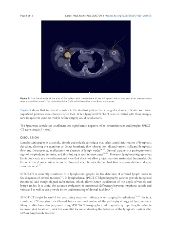
Figure 4. New lymph node at the arm of the patient with lymphedema of the left upper limb, at one year after lymphovenous
anastomosis (blue arrow). The contralateral side (right axilla) is marking a functional nodal group
Figure 5 shows that in patient number 8, the stardust pattern had changed and new reticular and linear
superficial patterns were observed after LVA. When lympho-SPECT/CT was correlated with these images,
new images that were not visible before surgery could be observed.
The Spearman correlation coefficient was significantly negative when circumferences and lympho-SPECT-
CT were tested (P = 0.02).
DISCUSSION
Lymphoscintigraphy is a specific, simple and reliable technique that offers useful information of lymphatic
function, allowing the examiner to detect lymphatic flow obstruction, dilated vessels, collateral lymphatic
flow and the presence, malfunction or absence of lymph nodes [8-10] . Dermal uptake is a pathognomonic
sign of lymphedema in limbs, and this finding is seen in most cases [11,12] . However, lymphoscintigraphy has
limitations such as a two-dimensional view that does not allow projection onto anatomical landmarks. On
the other hand, some artefacts can be observed when fibrosis, dermal backflow or accumulation in deeper
[13]
vessels is seen .
SPECT-CT is currently combined with lymphoscintigraphy for the detection of sentinel lymph nodes in
[14]
the diagnosis of several tumours . In lymphedema, SPECT-CT/lymphography systems provide integrated
functional and morphological information, which allows better localization of the depth of vessels and
lymph nodes. It is useful for accurate evaluation of anatomical differences between lymphatic vessels and
[15]
veins and as well, it can provide better understanding of dermal backflow .
SPECT-CT might be useful for predicting treatment efficacy when staging lymphedema [16-18] . In fact,
combined CT-imaging has allowed better comprehension of the pathophysiology of lymphedema.
Many studies have also proposed using SPECT-CT imaging beyond diagnosis by reporting its value in
microsurgical treatment, which is essential for understanding the behavior of the lymphatic system after
LVA or lymph node transfer.