
Page 85 - Read Online
P. 85
Page 6 of 12 Lasso. Plast Aesthet Res 2020;7:30 I http://dx.doi.org/10.20517/2347-9264.2019.75
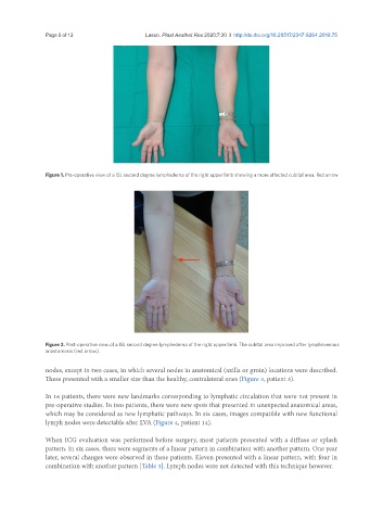
Figure 1. Pre-operative view of a ISL second degree lymphedema of the right upper limb showing a more affected cubital area. Red arrow
Figure 2. Post-operative view of a ISL second degree lymphedema of the right upper limb. The cubital area improved after lymphovenous
anastomosis (red arrow).
nodes, except in two cases, in which several nodes in anatomical (axilla or groin) locations were described.
These presented with a smaller size than the healthy, contralateral ones (Figure 3, patient 3).
In 16 patients, there were new landmarks corresponding to lymphatic circulation that were not present in
pre-operative studies. In two patients, there were new spots that presented in unexpected anatomical areas,
which may be considered as new lymphatic pathways. In six cases, images compatible with new functional
lymph nodes were detectable after LVA (Figure 4, patient 14).
When ICG evaluation was performed before surgery, most patients presented with a diffuse or splash
pattern. In six cases, there were segments of a linear pattern in combination with another pattern. One year
later, several changes were observed in these patients. Eleven presented with a linear pattern, with four in
combination with another pattern [Table 3]. Lymph nodes were not detected with this technique however.