
Page 135 - Read Online
P. 135
Page 8 of 13 Chao et al. Plast Aesthet Res. 2025;12:29 https://dx.doi.org/10.20517/2347-9264.2025.18
patients requires careful anterior dissection to avoid violating the prostatic capsule. Early recognition of
missteps allows for straightforward correction. Intrafascial dissection is also a valid alternative, connecting
perineal and robotic planes at the midline [16,41] . In our hands, however, infrafascial dissection enables rapid,
safe, and reproducible connection of the perineal and robotic planes, allowing blunt access to the lateral
urogenital triangle and tactile guidance for midline attachments, reducing the need for routine digital rectal
examination or specialized retractors and maintaining a low intraoperative injury rate [Table 1].
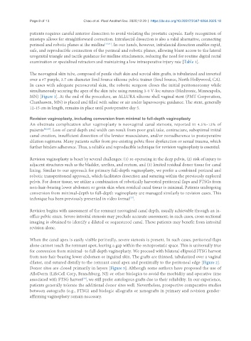
The neovaginal skin tube, composed of penile shaft skin and scrotal skin grafts, is tubularized and inverted
over a #7 purple, 3.7 cm-diameter Soul Source silicone pelvic trainer (Soul Source, North Hollywood, CA).
In cases with adequate penoscrotal skin, the robotic surgeon closes the initial peritoneotomy while
simultaneously securing the apex of the skin tube using running 3-0 V-loc sutures (Medtronic, Minneapolis,
MN) [Figure 1]. At the end of the procedure, an ALLURA silicone shell vaginal stent (PMT Corporation,
Chanhassen, MN) is placed and filled with saline or air under laparoscopic guidance. The stent, generally
12-15 cm in length, remains in place until postoperative day 5.
Revision vaginoplasty, including conversion from minimal to full-depth vaginoplasty
An obstinate complication after vaginoplasty is neovaginal canal stenosis, reported in 4.5%-12% of
patients [42,43] . Loss of canal depth and width can result from poor graft take, contracture, suboptimal initial
canal creation, insufficient dissection of the levator musculature, and/or nonadherence to postoperative
dilation regimens. Many patients suffer from pre-existing pelvic floor dysfunction or sexual trauma, which
further hinders adherence. Thus, a reliable and reproducible technique for revision vaginoplasty is essential.
Revision vaginoplasty is beset by several challenges: (1) re-operating in the deep pelvis, (2) risk of injury to
adjacent structures such as the bladder, urethra, and rectum, and (3) limited residual donor tissue for canal
lining. Similar to our approach for primary full-depth vaginoplasty, we prefer a combined perineal and
robotic transperitoneal approach, which facilitates dissection and suturing within the previously explored
pelvis. For donor tissue, we utilize a combination of robotically harvested peritoneal flaps and FTSGs from
non-hair-bearing lower abdomen or groin skin when residual canal tissue is minimal. Patients undergoing
conversion from minimal-depth to full-depth vaginoplasty are managed similarly to revision cases. This
technique has been previously presented in video format .
[37]
Revision begins with assessment of the remnant neovaginal canal depth, usually achievable through an in-
office pelvic exam. Severe introital stenosis may preclude accurate assessment; in such cases, cross-sectional
imaging is obtained to identify a dilated or sequestered canal. These patients may benefit from introital
revision alone.
When the canal apex is easily visible perineally, severe stenosis is present. In such cases, peritoneal flaps
alone cannot reach the remnant apex, leaving a gap within the rectoprostatic space. This is universally true
for conversion from minimal- to full-depth vaginoplasty. We proceed with bilateral ellipsoid FTSG harvest
from non-hair-bearing lower abdomen or inguinal skin. The grafts are thinned, tubularized over a vaginal
dilator, and sutured distally to the remnant canal apex and proximally to the peritoneal edge [Figure 2].
Donor sites are closed primarily in layers [Figure 3]. Although some authors have proposed the use of
AlloDerm (LifeCell Corp, Branchburg, NJ) or other biologics to avoid the morbidity and operative time
associated with FTSG harvest , we still prefer autologous grafts due to their reliability. In our experience,
[29]
patients generally tolerate the additional donor sites well. Nevertheless, prospective comparative studies
between autografts (e.g., FTSG) and biologic allografts or xenografts in primary and revision gender-
affirming vaginoplasty remain necessary.