
Page 59 - Read Online
P. 59
Horch et al. Plast Aesthet Res 2018;5:26 I http://dx.doi.org/10.20517/2347-9264.2018.25 Page 5 of 9
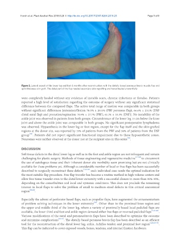
Figure 5. Lateral aspect of the lower leg and foot 6 months after reconstruction with the distally based peroneus brevis muscle flap and
split-thickness skin graft. The distal part of the flap needed secondary skin regrafting and hence healed uneventfully
were completely healed without any evidence of instable scars, chronic infections or fistulae. Patients
reported a high level of satisfaction regarding the outcome of surgery without any significant statistical
difference between the compared flaps. The active total range of motion was comparable in both groups
without significant differences [extension/flexion 78.5% ± 20.0% (PBF peroneus flap), 66.6% ± 23.1% (DSF
distal sural flap) and pronation/supination 70.9% ± 27.7% (PBF), 61.1% ± 33.3% (DSF)]. No instability of the
ankle joint was observed in patients from both groups. Circumference of the lower leg 15 cm below the knee
joint and above the ankle joint was comparable in both groups. No significant postoperative lymphedema
was observed. Hypaesthesia in the lower leg or foot region, except for the flap itself and the skin-grafted
regions at the donor site, was reported by 21% of patients from the PBF and 58% of patients from the DSF
[36]
group . Patients did not report significant functional impairment due to these hypoaesthetic zones.
[36]
Neuromas were neither observed at the donor nor at the recipient sites in this series .
DISCUSSION
Soft tissue defects in the distal lower leg as well as in the foot and ankle region are not infrequent and remain
challenging for plastic surgery. Methods of tissue engineering and regenerative medicine [37-51] to circumvent
the use of autologous tissue and their inherent donor site morbidity seem promising but are not clinically
available for these problems yet. Although a considerable number of local or free flaps has been successfully
described to surgically reconstruct these defects [7,52-58] each individual case needs the optimal indication for
the most suitable flap procedure. Free flap transfer has become a routine method in high volume centers and
allow free tissue transfer even to the distal lower extremity with a successful closure in more than 90%-95%,
depending on the comorbidities and local and systemic conditions. This does not preclude the remaining
interest in local flaps to solve the problem of small to medium-sized defects in this critical anatomical
region [59,60] .
Especially the advent of perforator based flaps, such as propeller flaps, have augmented the armamentarium
[52]
of problem solving techniques in the lower extremity . Other than in the proximal knee region and
the upper and middle third of the lower leg, where a variety of proximally based local pedicled flaps are
available, the lower third and foot and ankle region demand either free flaps or reversed pedicled flaps [11,61,62] .
Various modifications of the sural and peroneusbrevis flaps have been described to optimize the outcome
and minimize complications [63-67] . The distally based peroneus brevis flap has been described as an efficient
tool for the reconstruction of the distal lower leg, ankle, Achilles tendon and proximal foot region [4,65,68-73] .
This flap can be indicated to cover exposed vessels, bones, tendons, and internal fixation hardware.