
Page 57 - Read Online
P. 57
Horch et al. Plast Aesthet Res 2018;5:26 I http://dx.doi.org/10.20517/2347-9264.2018.25 Page 3 of 9
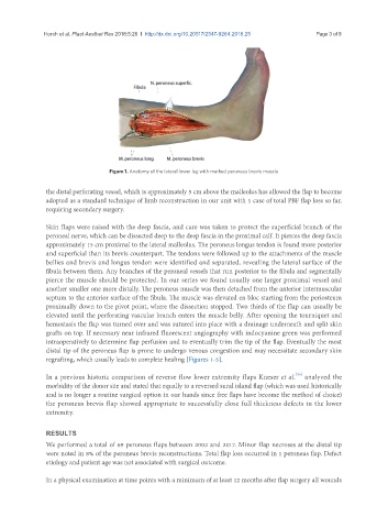
Figure 1. Anatomy of the lateral lower leg with marked peroneus brevis muscle
the distal perforating vessel, which is approximately 5 cm above the malleolus has allowed the flap to become
adopted as a standard technique of limb reconstruction in our unit with 1 case of total PBF flap loss so far,
requiring secondary surgery.
Skin flaps were raised with the deep fascia, and care was taken to protect the superficial branch of the
peroneal nerve, which can be dissected deep to the deep fascia in the proximal calf. It pierces the deep fascia
approximately 15 cm proximal to the lateral malleolus. The peroneus longus tendon is found more posterior
and superficial than its brevis counterpart. The tendons were followed up to the attachments of the muscle
bellies and brevis and longus tendon were identified and separated, revealing the lateral surface of the
fibula between them. Any branches of the peroneal vessels that run posterior to the fibula and segmentally
pierce the muscle should be protected. In our series we found usually one larger proximal vessel and
another smaller one more distally. The peroneus muscle was then detached from the anterior intermuscular
septum to the anterior surface of the fibula. The muscle was elevated en bloc starting from the periosteum
proximally down to the pivot point, where the dissection stopped. Two thirds of the flap can usually be
elevated until the perforating vascular branch enters the muscle belly. After opening the tourniquet and
hemostasis the flap was turned over and was sutured into place with a drainage underneath and split skin
grafts on top. If necessary near infrared fluorescent angiography with indocyanine green was performed
intraoperatively to determine flap perfusion and to eventually trim the tip of the flap. Eventually the most
distal tip of the peroneus flap is prone to undergo venous congestion and may necessitate secondary skin
regrafting, which usually leads to complete healing [Figures 1-5].
[36]
In a previous historic comparison of reverse flow lower extremity flaps Kneser et al. analyzed the
morbidity of the donor site and stated that equally to a reversed sural island flap (which was used historically
and is no longer a routine surgical option in our hands since free flaps have become the method of choice)
the peroneus brevis flap showed appropriate to successfully close full thickness defects in the lower
extremity.
RESULTS
We performed a total of 69 peroneus flaps between 2003 and 2017. Minor flap necroses at the distal tip
were noted in 8% of the peroneus brevis reconstructions. Total flap loss occurred in 1 peroneus flap. Defect
etiology and patient age was not associated with surgical outcome.
In a physical examination at time points with a minimum of at least 12 months after flap surgery all wounds