
Page 83 - Read Online
P. 83
Bolletta et al. Plast Aesthet Res 2019;6:22 I http://dx.doi.org/10.20517/2347-9264.2019.22 Page 9 of 14
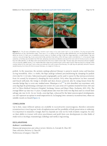
A B
C D
E F
Figure 3. A: The 25-year-old patient had a severe crush injury to his right lower limb in a car accident. The leg survived after
thrombectomy of the right femoral artery. There was a 12 cm defect of the right tibia after debridement, and the fractured fibula was
plated as shown; B: there was no available recipient artery in the thigh and leg. In the first stage operation, a radial forearm flap was
used as a vascular bridge flap, it was connected to the posterior tibial artery of the left leg in end-to-side fashion; C: in the second stage
operation, a free flap was harvested from the back, including myocutanous latissimus dorsi and the lower part of serratus muscle, carrying
two ribs (6th and 8th); D: the flaps were connected to the free end of radial forearm flap. The two legs were temporarily bound together
with an external skeletal fixator; E: four weeks later, the bridge was divided and part of the radial forearm flap was used for coverage of the
residual defect of the right leg; F: bone union was achieved and, with proper physiotherapy, the right leg was gradually trained to resume
weight-bearing. As shown, the ribs increased thickness, in a long term follow up
pedicle. In the meantime, the patients undergo physical therapy to preserve muscle status and function
during immobility. After 3-4 weeks, the flaps undergo ischemic preconditioning by clamping the pedicle
every day for 15 minutes. Indocyanine green angiography can be used to assess the flap neovascularization
from the wound, by temporarily clamping the main pedicle. Only when flap perfusion has been assessed
and found sufficient, the bridge is divided and skin closure achieved, also by using tissues from the
[122]
vascular bridge flap to cover any residual areas. Manrique et al. in 2018 described our experience with
cross-leg flaps by performing a retrospective review of a case series of 53 patients treated between 1985 and
2017 in China Medical University Hospital, Taichung, Taiwan and Mayo Clinic, Rochester, MN, USA. The
average follow-up time was 7.5 years. Complications rates were low (with two flap loss) and the overall limb
salvage rate was 96.2%. In our hands, cross-leg flaps, enhanced by the latest microsurgical developments,
can still represent an option to avoid amputation in challenging lower extremity reconstructions, where no
suitable vessels are found [Figure 3].
CONCLUSION
Up to date, many different options are available to reconstructive microsurgeons, therefore extremity
reconstruction is reaching new levels of sophistication and the possibility of limb preservation is widening.
It is important to remember, though, that this depends not only on the work of plastic surgeons, but also
on their ability to interact with other practitioners and profit form new developments in other fields of
study such as oncology, traumatology, radiology and medical engineering.
DECLARATIONS
Authors’ contributions
Manuscript preparation and critical review: Bolletta A, Corrado R, Chen HC
Data collection: Bolletta A, Chen HC
Performance of surgery: Chen HC