
Page 81 - Read Online
P. 81
Bolletta et al. Plast Aesthet Res 2019;6:22 I http://dx.doi.org/10.20517/2347-9264.2019.22 Page 7 of 14
A B
C D
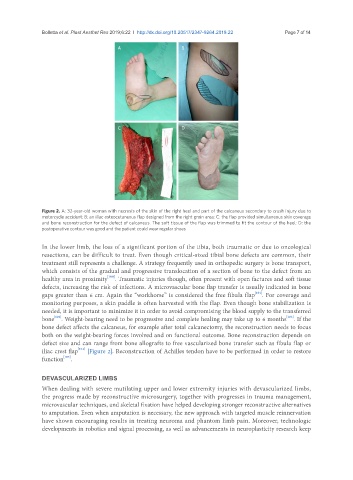
Figure 2. A: 32-year-old woman with necrosis of the skin of the right heel and part of the calcaneus secondary to crush injury due to
motorcycle accident; B: an iliac osteocutaneous flap designed from the right groin area; C: the flap provided simultaneous skin coverage
and bone reconstruction for the defect of calcaneus. The soft tissue of the flap was trimmed to fit the contour of the heel; D: the
postoperative contour was good and the patient could wear regular shoes
In the lower limb, the loss of a significant portion of the tibia, both traumatic or due to oncological
resections, can be difficult to treat. Even though critical-sized tibial bone defects are common, their
treatment still represents a challenge. A strategy frequently used in orthopedic surgery is bone transport,
which consists of the gradual and progressive translocation of a section of bone to the defect from an
[100]
healthy area in proximity . Traumatic injuries though, often present with open factures and soft tissue
defects, increasing the risk of infections. A microvascular bone flap transfer is usually indicated in bone
[101]
gaps greater than 6 cm. Again the “workhorse” is considered the free fibula flap . For coverage and
monitoring purposes, a skin paddle is often harvested with the flap. Even though bone stabilization is
needed, it is important to minimize it in order to avoid compromising the blood supply to the transferred
[102]
[103]
bone . Weight-bearing need to be progressive and complete healing may take up to 6 months . If the
bone defect affects the calcaneus, for example after total calcanectomy, the reconstruction needs to focus
both on the weight-bearing forces involved and on functional outcome. Bone reconstruction depends on
defect size and can range from bone allografts to free vascularized bone transfer such as fibula flap or
[104]
iliac crest flap [Figure 2]. Reconstruction of Achilles tendon have to be performed in order to restore
[105]
function .
DEVASCULARIZED LIMBS
When dealing with severe mutilating upper and lower extremity injuries with devascularized limbs,
the progress made by reconstructive microsurgery, together with progresses in trauma management,
microvascular techniques, and skeletal fixation have helped developing stronger reconstructive alternatives
to amputation. Even when amputation is necessary, the new approach with targeted muscle reinnervation
have shown encouraging results in treating neuroma and phantom limb pain. Moreover, technologic
developments in robotics and signal processing, as well as advancements in neuroplasticity research keep