
Page 50 - Read Online
P. 50
Page 8 of 12 Wee et al. Plast Aesthet Res 2019;6:12 I http://dx.doi.org/10.20517/2347-9264.2019.02
Figure 9. Radiograph demonstrating tibial nonunion despite previous fixation and allografting attempts
A B
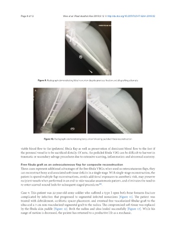
Figure 10. Radiographs demonstrating bony union following pedicled fibula reconstruction
viable blood flow to the ipsilateral fibula flap as well as preservation of dominant blood flow to the foot if
the peroneal vessel is to be sacrificed distally. Of note, the pedicled fibula VBG can be difficult to harvest in
traumatic or secondary salvage procedures due to extensive scarring, inflammation and abnormal anatomy.
Free fibula graft as an osteocutaneous flap for composite reconstruction
These cases represent additional advantages of the free fibula VBGs; when used as osteocutaneous flaps, they
can reconstruct bony and associated soft tissue deficits in a single stage. With single-stage reconstruction, the
patient is spared multiple flap reconstructions, avoids additional exposures to anesthetic risk, may preserve
recipient vessels when performed in an end-to-side vascular anastomosis pattern, and eliminates the need to
re-enter scarred wound beds for subsequent staged procedures .
[20]
Case 9. This patient was 22-year-old army soldier who suffered a type I open both-bone forearm fracture
complicated by infection that progressed to segmental infected nonunions [Figure 11]. The patient was
treated with debridement, antibiotic spacer placement, and eventual free vascularized fibular graft to the
ulna and a 3 cm non-vascularized segmental graft to the radius. The compromised soft tissue was replaced
by the fibula skin paddle [Figure 12]. Both the radius and ulna healed successfully [Figure 13]. While his
range of motion is decreased, the patient has returned to a productive life as a mechanic.