
Page 49 - Read Online
P. 49
Wee et al. Plast Aesthet Res 2019;6:12 I http://dx.doi.org/10.20517/2347-9264.2019.02 Page 7 of 12
Figure 7. Free fibula used in conjunction with allograft, per the Capanna technique
A B C
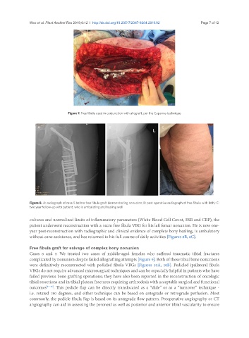
Figure 8. A: radiograph of case 5 before free fibula graft demonstrating nonunion; B: post operative radiograph of free fibula with IMN; C:
two year follow-up with patient, who is ambulating and healing well
cultures and normalized limits of inflammatory parameters (White Blood Cell Count, ESR and CRP), the
patient underwent reconstruction with a 14cm free fibula VBG for his left femur nonunion. He is now one-
year post-reconstruction with radiographic and clinical evidence of complete bony healing, is ambulatory
without cane assistance, and has returned to his full course of daily activities [Figures 8B, 8C].
Free fibula graft for salvage of complex bony nonunion
Cases 6 and 7. We treated two cases of middle-aged females who suffered traumatic tibial fractures
complicated by nonunion despite failed allografting attempts [Figure 9]. Both of these tibial bone nonunions
were definitively reconstructed with pedicled fibula VBGs [Figures 10A, 10B]. Pedicled ipsilateral fibula
VBGs do not require advanced microsurgical techniques and can be especially helpful in patients who have
failed previous bone grafting operations; they have also been reported in the reconstruction of oncologic
tibial resections and in tibial plateau fractures requiring arthrodesis with acceptable surgical and functional
outcomes [17-19] . This pedicle flap can be directly translocated as a “slide” or as a “turnover” technique -
i.e. rotated 180 degrees, and either technique can be based on antegrade or retrograde perfusion. Most
commonly, the pedicle fibula flap is based on its antegrade flow pattern. Preoperative angiography or CT
angiography can aid in assessing the peroneal as well as posterior and anterior tibial vascularity to ensure