
Page 130 - Read Online
P. 130
Page 8 of 11 Zhang et al. Plast Aesthet Res 2019;6:30 I http://dx.doi.org/10.20517/2347-9264.2019.040
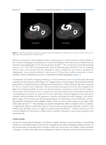
Figure 3. Computed tomographic angiography imaging of traumatized extremity with identification of potential recipient vessel prior to
planned perforator flap for anterior tibial soft tissue
However, preoperative vessel imaging remains common practice in the traumatic setting. Similar to
the evolution of imaging for the purposes of injury identification, vessel selection has evolved from the
transition from angiography to CTA over the previous decade [64,65] . For the purposes of free-flap planning,
[66]
Duymaz et al. were able to demonstrate the utility of obtaining lower extremity CTAs in correlating
arterial injury with eventual flap loss, although no direct comparisons were made to preoperative
angiography. As previously mentioned, routine use of CTA provides excellent assessment of lower-
extremity anatomy without the associated co-morbidities of formal angiography [Figure 3].
Development of ancillary imaging modalities to assess perforator vessel for preoperative planning
[67]
continues to evolve. Recent work by Feng et al. suggests the use of color doppler ultrasound demonstrates
greater fidelity of identifying and localizing dominant perforators of lower extremity flap when compared
to CTA in a head-to-head comparison. The use of Indocyanine green (ICG) has also emerged as an
adjunctive imaging modality to assess the microvasculature of perforator and local tissue flaps in
microsurgical reconstruction. ICG is a cyanine dye with near-infrared spectral absorbance that binds
circulating plasma proteins. As such, ICG in concert with near infrared imaging has been used across
multiple medical disciplines for the purposes of vascular and lymph perfusion imaging. In the field of
plastic surgery, ICG has been used with increasing frequency for the purposes of local, perforator, and free-
flap perfusion distribution. Most published studies to date use ICG to assess viability of skin flaps of the
trunk, head, and neck [68-70] . The technology was recently demonstrated, albeit in a limited series of 23 patients,
to significantly improve complication rates of tissue necrosis and deep-space infection in patients with
[71]
Gustilo Type IIIB when used as an adjunct to guide initial debridement . While its use has yet to be
routinely adopted, ICG has proven a reliable adjunct available to clinicians to assess tissue perfusion in the
operating room.
CONCLUSION
Advances in microsurgical techniques, the advent of negative pressure wound technology in temporizing
wound care, and improvements in preoperative imaging have facilitated changing treatment practices in
the reconstruction of traumatic lower extremity injuries over the previous two decades. Despite persistent
challenges, as evidenced by high rates of postoperative infection, flap loss, and poor functional recovery,