
Page 129 - Read Online
P. 129
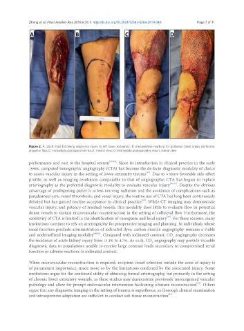
Zhang et al. Plast Aesthet Res 2019;6:30  I  http://dx.doi.org/10.20517/2347-9264.2019.040                                        Page 7 of 11


               Figure 2. A: adult male following degloving injury to left lower extremity; B: preoperative marking for posterior tibial artery perforator
               propeller flap; C: immediate postoperative result, medial view; D: immediate postoperative result, lateral view


               performance and cost to the hospital system [53,54] . Since its introduction in clinical practice in the early
               1990s, computed tomographic angiography (CTA) has become the de-facto diagnostic modality of choice
                                                                        [55]
               to assess vascular injury in the setting of lower-extremity trauma . Due to a more favorable side-effect
               profile, as well as imaging resolution comparable to that of angiography, CTA has begun to replace
               arteriography as the preferred diagnostic modality to evaluate vascular injury [56,57] . Despite the obvious
               advantage of predisposing patient’s to less ionizing radiation and the avoidance of complications such as
               pseudoaneurysm, vessel thrombosis, and vessel injury, the routine use of CTA has long been continuously
                                                                      [58]
               debated but has gained routine acceptance in clinical practice . While CT imaging may demonstrate
               vascular injury, and patency of residual vessels, this modality does little to evaluate flow in potential
               donor vessels to sustain microvascular reconstruction in the setting of collateral flow. Furthermore, the
                                                                                  [59]
               sensitivity of CTA is limited in the identification of vasospasm and local injury . For these reasons, many
               institutions continue to rely on arteriography for preoperative imaging and planning. In individuals whose
               renal function preclude administration of iodinated dyes, carbon dioxide angiography remains a viable
               and underutilized imaging modality [60,61] . Compared with iodinated contrast, CO  angiography decreases
                                                                                     2
               the incidence of acute kidney injury from 11.1% to 4.7%. As such, CO  angiography may provide valuable
                                                                           2
               diagnostic data in populations unable to receive large contrast loads secondary to compromised renal
               function or adverse reactions to iodinated contrast.

               When microvascular reconstruction is required, recipient vessel selection outside the zone of injury is
               of paramount importance, made more so by the limitations conferred by the associated injury. Some
               institutions argue for the continued utility of obtaining formal arteriography, but primarily in the setting
               of chronic lower extremity wounds, as these studies may demonstrate previously unrecognized vascular
                                                                                                 [62]
               pathology and allow for prompt endovascular intervention facilitating ultimate reconstruction . Others
               argue that any diagnostic imaging in the setting of trauma is superfluous, as thorough clinical examination
                                                                                   [63]
               and intraoperative adaptation are sufficient to conduct soft-tissue reconstruction .