
Page 114 - Read Online
P. 114
Cohen-Shohet et al. Plast Aesthet Res 2019;5:28 I http://dx.doi.org/10.20517/2347-9264.2019.030 Page 5 of 13
Figure 3. One-month post-operation: There is good healing over the wound without any distal necrosis
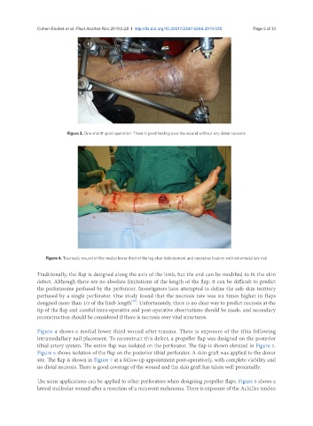
Figure 4. Traumatic wound of the medial lower third of the leg after debridement and operative fixation with intramedullary nail
Traditionally, the flap is designed along the axis of the limb, but the end can be modified to fit the skin
defect. Although there are no absolute limitations of the length of the flap, it can be difficult to predict
the perforasome perfused by the perforator. Investigators have attempted to define the safe skin territory
perfused by a single perforator. One study found that the necrosis rate was six times higher in flaps
[25]
designed more than 1/3 of the limb length . Unfortunately, there is no clear way to predict necrosis at the
tip of the flap and careful intra-operative and post-operative observations should be made, and secondary
reconstruction should be considered if there is necrosis over vital structures.
Figure 4 shows a medial lower third wound after trauma. There is exposure of the tibia following
intramedullary nail placement. To reconstruct this defect, a propeller flap was designed on the posterior
tibial artery system. The entire flap was isolated on the perforator. The flap is shown elevated in Figure 5.
Figure 6 shows isolation of the flap on the posterior tibial perforator. A skin graft was applied to the donor
site. The flap is shown in Figure 7 at a follow-up appointment post-operatively, with complete viability and
no distal necrosis. There is good coverage of the wound and the skin graft has taken well proximally.
The same applications can be applied to other perforators when designing propeller flaps. Figure 8 shows a
lateral malleolar wound after a resection of a recurrent melanoma. There is exposure of the Achilles tendon