
Page 119 - Read Online
P. 119
Page 10 of 13 Cohen-Shohet et al. Plast Aesthet Res 2019;5:28 I http://dx.doi.org/10.20517/2347-9264.2019.030
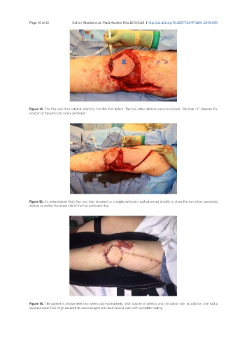
Figure 14. The flap was then rotated inferiorly into the first defect. The two other defects were connected. The blue “X” denotes the
location of the genicular artery perforator
Figure 15. An anterolateral thigh flap was then elevated on a single perforator and advanced distally to close the two other connected
defects as well as the donor site of the first perforator flap
Figure 16. The patient is shown here two weeks post-operatively, with closure of defects and the donor site. In addition, she had a
separate superficial thigh wound that was managed with local wound care, with complete healing