
Page 89 - Read Online
P. 89
Page 4 of 10 Tuinder et al. Plast Aesthet Res 2024;11:38 https://dx.doi.org/10.20517/2347-9264.2024.40
Figure 1. An example of preoperative markings at the donor site for the lateral thigh perforator (LTP) flap.
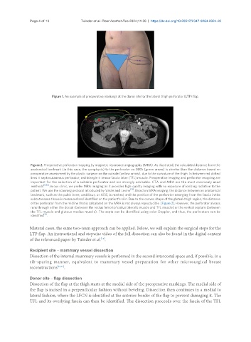
Figure 2. Preoperative perforator mapping by magnetic resonance angiography (MRA). As illustrated, the calculated distance from the
anatomical landmark (in this case, the symphysis) to the perforator on MRA (green arrow) is shorter than the distance based on
preoperative assessment by the plastic surgeon on the outside (yellow arrow), due to the curvature of the thigh. In between red dotted
lines = septocutaneous perforator; red triangle = tensor fascia latae (TFL) muscle. Preoperative imaging and perforator mapping are
important for the selection of a suitable perforator and are strongly advisable. CTA and MRA are the most commonly used
methods [8,10] . In our clinic, we prefer MRA imaging as it provides high-quality imaging with no exposure of ionizing radiation to the
patient. We use the scanning protocol introduced by Vasile and Levine [13] . Based on MRA imaging, the distance between an anatomical
landmark, such as the pubic bone, umbilicus, or ASIS, is marked, and the position of the perforator emerging from the fascia in the
subcutaneous tissue is measured and identified on the patient’s skin. Due to the convex shape of the gluteal-thigh region, the distance
of the perforator from the midline that is calculated on the MRA is not always reproducible [Figure 2]. However, the perforator always
runs through either the dorsal (between the rectus femoris/vastus lateralis muscle and TFL muscle) or the ventral septum (between
the TFL muscle and gluteus medius muscle). The septa can be identified using color Doppler, and thus, the perforators can be
[8]
identified .
bilateral cases, the same two-team approach can be applied. Below, we will explain the surgical steps for the
LTP flap. An instructional and stepwise video of the full dissection can also be found in the digital content
of the referenced paper by Tuinder et al. .
[15]
Recipient site – mammary vessel dissection
Dissection of the internal mammary vessels is performed in the second intercostal space and, if possible, in a
rib-sparing manner, equivalent to mammary vessel preparation for other microsurgical breast
reconstructions [16,17] .
Donor site – flap dissection
Dissection of the flap at the thigh starts at the medial side of the preoperative markings. The medial side of
the flap is incised in a perpendicular fashion without beveling. Dissection then continues in a medial to
lateral fashion, where the LFCN is identified at the anterior border of the flap to prevent damaging it. The
TFL and its overlying fascia can then be identified. The dissection proceeds over the fascia of the TFL