
Page 91 - Read Online
P. 91
Page 6 of 10 Tuinder et al. Plast Aesthet Res 2024;11:38 https://dx.doi.org/10.20517/2347-9264.2024.40
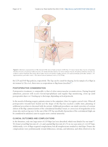
Figure 3. Schematic representation of the harvest of the lateral thigh perforator (LTP) flap: (1) tensor fascia latae muscle; (2) gluteus
maximus muscle; (3) gluteus medius muscle; (4) vastus lateralis muscle; (5) rectus femoris muscle; (6) gluteus minimus muscle; (7)
posterior septum between the tensor fascia latae muscle and gluteus medius muscle; (8) septocutaneous perforator (artery) ; (9)
septocutaneous perforator (vein) ; (10) lateral femoral cutaneous nerve; (11) LTP flap.
the LTP and PAP flap pedicles, respectively. The flap can be coned for shaping. In the sample of 23 flaps in
six women by Chu et al., there were no reoperations or flap losses described .
[20]
POSTOPERATIVE CONSIDERATION
Postoperative treatment is comparable to that of other microvascular reconstructions. During hospital
admission, patients will receive thromboprophylaxis and regular flap monitoring, often up until
postoperative days 4 or 5 leading up to discharge, depending on local protocols.
In the months following surgery, patients return to the outpatient clinic for regular control visits. When all
postoperative wounds have healed and the shape of the flap has reached a stable state, planning of
additional procedures is discussed with the patient. Additional procedures can entail correction of contour
defects of the flap, symmetrization of the contralateral (healthy) breast, or correction of irregularities at the
donor site, such as dog ears. In the case of the LTP flap specifically, liposuction of the contralateral thigh can
be considered in unilateral cases to acquire more contour symmetry.
CLINICAL OUTCOMES AND COMPLICATIONS
In the literature, only one large series of LTP flaps has been described, which was detailed by our team .
[15]
We found a total flap loss rate of 1.4% and a partial flap loss rate of 0.7% in our case series of 138 LTP flaps.
Additionally, 8.0% of flaps required reexploration, but all reexplorations resulted in vital flaps. Donor site
complications were predominantly wound dehiscence, seroma, and infection, and often observed in the