
Page 81 - Read Online
P. 81
Page 6 of 10 Hammond et al. Plast Aesthet Res 2024;11:28 https://dx.doi.org/10.20517/2347-9264.2024.27
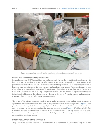
Figure 4. Intraoperative patient and robotic arm positioning during robotic latissimus muscle flap harvest.
Robotic deep inferior epigastric perforator flap
Standard abdominal DIEP flap markings are used preoperatively, and the patient is positioned supine with
bilateral arms abducted on arm boards. The operation begins as a standard DIEP flap harvest until
perforators are isolated and chosen. Anterior dissection of the perforator under loupe magnification is
limited to only where the perforator exits the inner surface of the rectus muscle. Pneumoperitoneum is then
obtained to 15 mmHg utilizing Varess needle insufflation. Three robot ports are then placed through the
fascia on the contralateral side of the flap along the anterior axillary line. The robot is positioned at bedside
to the ipsilateral flap, and the robotic arms are docked to the ports. A bipolar grasper and monopolar
scissors are then introduced under endoscopic visualization.
The course of the inferior epigastric vessels is traced under endoscopic vision, and the posterior sheath is
opened to facilitate circumferential dissection of the pedicle free from surrounding tissues [Figure 6]. The
pedicle is then clipped, divided, and pulled gently through the small anterior facial defect. A barbed suture is
then introduced into the abdomen and used to close the posterior sheath [Figure 7]. If a bilateral DIEP flap
is planned, the robot can be rotated for harvest on the contralateral side. After flap harvest and extraction,
the ports are removed, and port sites are closed. DIEP flap inset and microsurgical anastomosis are then
performed in a traditional fashion.
POSTOPERATIVE CONSIDERATIONS
The postoperative approaches for robotic latissimus muscle flap and DIEP flap patients can vary and should