
Page 79 - Read Online
P. 79
Page 4 of 10 Hammond et al. Plast Aesthet Res 2024;11:28 https://dx.doi.org/10.20517/2347-9264.2024.27
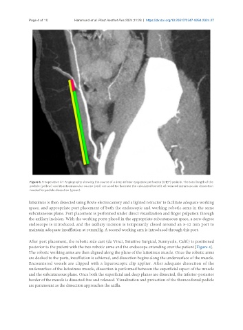
Figure 1. Preoperative CT Angiography showing the course of a deep inferior epigastric perforator (DIEP) pedicle. The total length of the
pedicle (yellow) and its intramuscular course (red) are used to illustrate the calculated benefit of reduced intramuscular dissection
needed for pedicle dissection (green).
latissimus is then dissected using Bovie electrocautery and a lighted retractor to facilitate adequate working
space, and appropriate port placement of both the endoscopic and working robotic arms in the same
subcutaneous plane. Port placement is performed under direct visualization and finger palpation through
the axillary incision. With the working ports placed in the appropriate subcutaneous space, a zero-degree
endoscope is introduced, and the axillary incision is temporarily closed around an 8-12 mm port to
maintain adequate insufflation at 10mmHg. A second working arm is introduced through this port.
After port placement, the robotic side cart (da Vinci, Intuitive Surgical, Sunnyvale, Calif.) is positioned
posterior to the patient with the two robotic arms and the endoscope extending over the patient [Figure 4].
The robotic working arms are then aligned along the plane of the latissimus muscle. Once the robotic arms
are docked to the ports, insufflation is achieved, and dissection begins along the undersurface of the muscle.
Encountered vessels are clipped with a laparoscopic clip applier. After adequate dissection of the
undersurface of the latissimus muscle, dissection is performed between the superficial aspect of the muscle
and the subcutaneous plane. Once both the superficial and deep planes are dissected, the inferior-posterior
border of the muscle is dissected free and released. Visualization and protection of the thoracodorsal pedicle
are paramount as the dissection approaches the axilla.