
Page 72 - Read Online
P. 72
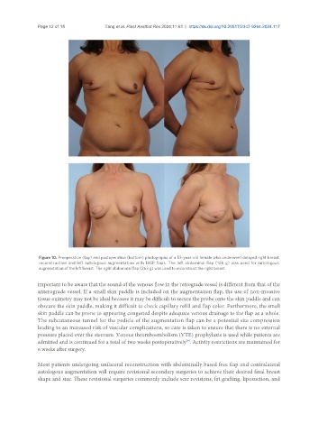
Page 12 of 15            Tang et al. Plast Aesthet Res 2024;11:61  https://dx.doi.org/10.20517/2347-9264.2024.117


                Figure 10. Preoperative (top) and postoperative (bottom) photographs of a 53-year-old female who underwent delayed right breast
                reconstruction and left autologous augmentation with DIEP flaps. The left abdominal flap (126 g) was used for autologous
                augmentation of the left breast. The right abdominal flap (263 g) was used to reconstruct the right breast.


               important to be aware that the sound of the venous flow in the retrograde vessel is different from that of the
               anterograde vessel. If a small skin paddle is included on the augmentation flap, the use of non-invasive
               tissue oximetry may not be ideal because it may be difficult to secure the probe onto the skin paddle and can
               obscure the skin paddle, making it difficult to check capillary refill and flap color. Furthermore, the small
               skin paddle can be prone to appearing congested despite adequate venous drainage to the flap as a whole.
               The subcutaneous tunnel for the pedicle of the augmentation flap can be a potential site compression
               leading to an increased risk of vascular complications, so care is taken to ensure that there is no external
               pressure placed over the sternum. Venous thromboembolism (VTE) prophylaxis is used while patients are
               admitted and is continued for a total of two weeks postoperatively . Activity restrictions are maintained for
                                                                       [9]
               6 weeks after surgery.

               Most patients undergoing unilateral reconstruction with abdominally based free flap and contralateral
               autologous augmentation will require revisional secondary surgeries to achieve their desired final breast
               shape and size. These revisional surgeries commonly include scar revisions, fat grafting, liposuction, and