
Page 66 - Read Online
P. 66
Page 6 of 15 Tang et al. Plast Aesthet Res 2024;11:61 https://dx.doi.org/10.20517/2347-9264.2024.117
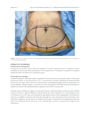
Figure 3. Flap marking includes a vertical line indicating the planned divide between the larger and the small flaps based on preoperative
CT scans/volumetric analysis.
OPERATIVE TECHNIQUE
Positioning and preparation
The patient is positioned supine with arms abducted. The bed is typically turned 180 degrees to allow
simultaneous access to the chest and abdomen for the surgical teams. The patient is prepped in the standard
fashion from the neck down to the suprapubic region.
Flap design and elevation
Standard marking for DIEP flap surgery is marked out over the abdomen, ensuring the capture of the major
perforators shown on the preoperative CTA. A vertical line is marked, indicating the planned divide
between the two flaps and is based on the ideal position for dividing the flaps based on preoperative CT
scans/volumetric analysis [Figure 3]. Incisions are made and standard DIEP flap elevation from lateral to
medial is performed. The superficial inferior epigastric veins (SIEV) are preserved.
Typically, flaps of different weights are used and division is performed based on the previously defined
location of the left or right hemiabdomen, determined by the volume needed for each breast [Figures 4 and
5]. Perfusion zones of the flaps can be assessed through physical exam or laser angiography. Once perforator
selection is established, intramuscular dissection through the rectus abdominus muscle and down to the
deep inferior epigastric vessels deep to the muscles is performed. The pedicle for the augmentation flap may
need to be dissected all the way down to the external iliac vessels to maximize the length for the
anastomosis.