
Page 11 - Read Online
P. 11
Gunderson et al. Plast Aesthet Res 2023;10:50 https://dx.doi.org/10.20517/2347-9264.2023.42 Page 5 of 15
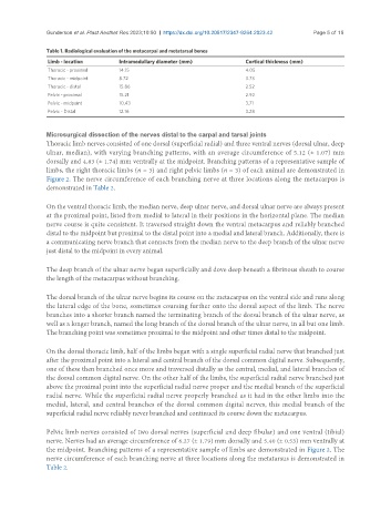
Table 1. Radiological evaluation of the metacarpal and metatarsal bones
Limb - location Intramedullary diameter (mm) Cortical thickness (mm)
Thoracic - proximal 14.15 4.05
Thoracic - midpoint 8.72 3.73
Thoracic - distal 15.86 2.52
Pelvic - proximal 15.21 2.92
Pelvic - midpoint 10.43 3.71
Pelvic - Distal 12.16 3.28
Microsurgical dissection of the nerves distal to the carpal and tarsal joints
Thoracic limb nerves consisted of one dorsal (superficial radial) and three ventral nerves (dorsal ulnar, deep
ulnar, median), with varying branching patterns, with an average circumference of 5.12 (± 1.07) mm
dorsally and 4.83 (± 1.74) mm ventrally at the midpoint. Branching patterns of a representative sample of
limbs, the right thoracic limbs (n = 3) and right pelvic limbs (n = 3) of each animal are demonstrated in
Figure 2. The nerve circumference of each branching nerve at three locations along the metacarpus is
demonstrated in Table 2.
On the ventral thoracic limb, the median nerve, deep ulnar nerve, and dorsal ulnar nerve are always present
at the proximal point, listed from medial to lateral in their positions in the horizontal plane. The median
nerve course is quite consistent. It traversed straight down the ventral metacarpus and reliably branched
distal to the midpoint but proximal to the distal point into a medial and lateral branch. Additionally, there is
a communicating nerve branch that connects from the median nerve to the deep branch of the ulnar nerve
just distal to the midpoint in every animal.
The deep branch of the ulnar nerve began superficially and dove deep beneath a fibrinous sheath to course
the length of the metacarpus without branching.
The dorsal branch of the ulnar nerve begins its course on the metacarpus on the ventral side and runs along
the lateral edge of the bone, sometimes coursing further onto the dorsal aspect of the limb. The nerve
branches into a shorter branch named the terminating branch of the dorsal branch of the ulnar nerve, as
well as a longer branch, named the long branch of the dorsal branch of the ulnar nerve, in all but one limb.
The branching point was sometimes proximal to the midpoint and other times distal to the midpoint.
On the dorsal thoracic limb, half of the limbs began with a single superficial radial nerve that branched just
after the proximal point into a lateral and central branch of the dorsal common digital nerve. Subsequently,
one of these then branched once more and traversed distally as the central, medial, and lateral branches of
the dorsal common digital nerve. On the other half of the limbs, the superficial radial nerve branched just
above the proximal point into the superficial radial nerve proper and the medial branch of the superficial
radial nerve. While the superficial radial nerve properly branched as it had in the other limbs into the
medial, lateral, and central branches of the dorsal common digital nerves, this medial branch of the
superficial radial nerve reliably never branched and continued its course down the metacarpus.
Pelvic limb nerves consisted of two dorsal nerves (superficial and deep fibular) and one ventral (tibial)
nerve. Nerves had an average circumference of 6.27 (± 1.79) mm dorsally and 5.40 (± 0.53) mm ventrally at
the midpoint. Branching patterns of a representative sample of limbs are demonstrated in Figure 2. The
nerve circumference of each branching nerve at three locations along the metatarsus is demonstrated in
Table 2.