
Page 95 - Read Online
P. 95
Myers et al. Plast Aesthet Res 2023;10:38 https://dx.doi.org/10.20517/2347-9264.2022.150 Page 7 of 13
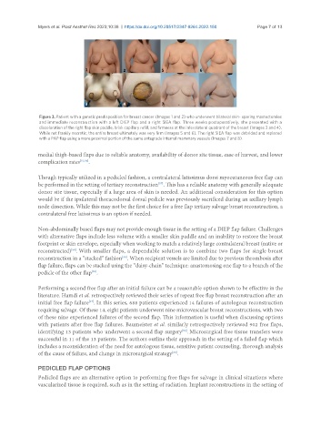
Figure 3. Patient with a genetic predisposition for breast cancer (Images 1 and 2) who underwent bilateral skin- sparing mastectomies
and immediate reconstruction with a left DIEP flap and a right SIEA flap. Three weeks postoperatively, she presented with a
discoloration of the right flap skin paddle, brisk capillary refill, and firmness at the inferolateral quadrant of the breast (Images 3 and 4).
While not frankly necrotic, the entire breast ultimately was very firm (Images 5 and 6). The right SIEA flap was debrided and replaced
with a PAP flap using a more proximal portion of the same antegrade internal mammary vessels (Images 7 and 8).
medial thigh-based flaps due to reliable anatomy, availability of donor site tissue, ease of harvest, and lower
complication rates [53,56] .
Though typically utilized in a pedicled fashion, a contralateral latissimus dorsi myocutaneous free flap can
[57]
be performed in the setting of tertiary reconstruction . This has a reliable anatomy with generally adequate
donor site tissue, especially if a large area of skin is needed. An additional consideration for this option
would be if the ipsilateral thoracodorsal dorsal pedicle was previously sacrificed during an axillary lymph
node dissection. While this may not be the first choice for a free flap tertiary salvage breast reconstruction, a
contralateral free latissimus is an option if needed.
Non-abdominally based flaps may not provide enough tissue in the setting of a DIEP flap failure. Challenges
with alternative flaps include less volume with a smaller skin paddle and an inability to restore the breast
footprint or skin envelope, especially when working to match a relatively large contralateral breast (native or
reconstructed) . With smaller flaps, a dependable solution is to combine two flaps for single breast
[58]
[39]
reconstruction in a “stacked” fashion . When recipient vessels are limited due to previous thrombosis after
flap failure, flaps can be stacked using the “daisy-chain” technique: anastomosing one flap to a branch of the
pedicle of the other flap .
[59]
Performing a second free flap after an initial failure can be a reasonable option shown to be effective in the
literature. Hamdi et al. retrospectively reviewed their series of repeat free flap breast reconstruction after an
[17]
initial free flap failure . In this series, 688 patients experienced 14 failures of autologous reconstruction
requiring salvage. Of these 14, eight patients underwent nine microvascular breast reconstructions, with two
of these nine experienced failures of the second flap. This information is useful when discussing options
with patients after free flap failures. Baumeister et al. similarly retrospectively reviewed 902 free flaps,
identifying 13 patients who underwent a second flap surgery . Microsurgical free tissue transfers were
[60]
successful in 11 of the 13 patients. The authors outline their approach in the setting of a failed flap which
includes a reconsideration of the need for autologous tissue, sensitive patient counseling, thorough analysis
[60]
of the cause of failure, and change in microsurgical strategy .
PEDICLED FLAP OPTIONS
Pedicled flaps are an alternative option to performing free flaps for salvage in clinical situations where
vascularized tissue is required, such as in the setting of radiation. Implant reconstructions in the setting of