
Page 94 - Read Online
P. 94
Page 6 of 13 Myers et al. Plast Aesthet Res 2023;10:38 https://dx.doi.org/10.20517/2347-9264.2022.150
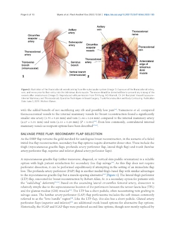
Figure 2. Illustration of the thoracodorsal vessels arising from the subscapular system (Image 1). Exposure of the thoracodorsal artery,
vein, and nerve prior to their entry into the latissimus dorsi muscle. The nerve should be dissected free to prevent any kinking of the
vessels after anastomosis (Image 2). Reproduced with permission from TA Kung, AO Momoh, Ch 24: Recipient Vessel Exposure--
Internal Mammary and Thoracodorsal, Operative Techniques in Breast Surgery, Trunk Reconstruction and Body Contouring. Publication
Date: June 3, 2019. Wolters Kluwer.
with the added benefit of not sacrificing any rib and possibly less pain . Yamamoto et al. compared
[46]
thoracoacromial vessels to the internal mammary vessels for breast reconstruction found a significantly
smaller size artery (1.70 ± 0.26 mm) and vein (1.64 ± 0.24 mm) compared to the internal mammary artery
(2.27 ± 0.31 mm) and vein (2.33 ± 0.29 mm) (P < 0.001) . Even less commonly, contralateral internal
[46]
mammary vessels as recipient options have been described [47,48] .
SALVAGE FREE FLAP: SECONDARY FLAP SELECTION
As the DIEP flap remains the gold standard for autologous breast reconstruction, in the scenario of a failed
initial free flap reconstruction, secondary free flap options require alternative donor sites. These include the
thigh (myocutaneous gracilis flaps, profunda artery perforator flap, lateral thigh flap) and trunk (lumbar
artery perforator flap, superior and inferior gluteal artery perforator flaps).
A myocutaneous gracilis flap (either transverse, diagonal, or vertical skin paddle orientation) is a reliable
option with high patient satisfaction for secondary free flap salvage . As this flap does not require
[49]
perforator dissection, it can be performed expeditiously if attempting in the setting of an immediate flap
loss. The profunda artery perforator (PAP) flap is another medial thigh-based flap with similar advantages
[50]
to the myocutaneous gracilis flap but a muscle-sparing alternative [Figure 3]. The lateral thigh perforator
(LTP) flap, renovated for breast reconstruction by Robert Allen, Sr. is a secondary option for patients with
the “saddlebag” deformity [51,52] . Based on the ascending lateral circumflex femoral artery, dissection is
relatively simple due to the septocutaneous location of the perforators between the tensor fascia lata (TFL)
and the gluteus medius (GM) muscles . The LTP has a short pedicle, often necessitating vein grafting in
[53]
salvage cases. The lumbar artery perforator (LAP) flap perforasome includes the soft tissues commonly
referred to as the “love handle” region . Like the LTP flap, this also has a short pedicle. Gluteal artery
[54]
perforator flaps (superior and inferior) are additional trunk-based options for alternative flap options.
[55]
Historically, the SGAP and IGAP flaps were preferred second-line options, though now mostly replaced by