
Page 75 - Read Online
P. 75
Page 6 of 9 Black et al. Plast Aesthet Res 2023;10:31 https://dx.doi.org/10.20517/2347-9264.2023.04
2
[13]
without acellular dermal templates when the incidence rate of skin loss exceeding 10 cm surpassed 25.3% .
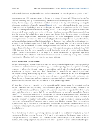
At our institution, DIEP reconstruction is performed in two stages following all NSM approaches: the first
involves harvesting the flap and anastomosing it to the internal mammary vessels in a standard fashion.
During the first stage, a large elliptical skin paddle is preserved on the flap for both banking and Doppler
ultrasound monitoring of vascular patency [Figure 2]. After two weeks (ample time for skin necrosis
demarcation and final pathology), we return to the operating room to completely remove the DIEP flap skin
- both the banked portion as well as the monitoring paddle - if tumor margins are negative or if there is no
skin necrosis. If tumor margins are positive or if there are significant amounts of full-thickness mastectomy
skin flap necrosis, the banked skin is used to reconstruct the skin defect due to oncologic re-excision or
necrosis. This technique provides a plan to manage necrosis and allows for improved cosmesis. Although a
second procedure is not without its risks, such as flap hypoperfusion during induction of general anesthesia,
we have not had any instances of flap failure attributed to the second stage banked skin excision in our ten-
year experience. Standardizing these methods for all patients undergoing NSM can lead to increased patient
satisfaction, cost-effectiveness, and overall stronger reconstructive outcomes. We have found that we use
banked skin in 18% of cases: 15% from skin necrosis and 3% from positive margins on final pathology. With
NSM, if the skin from the areola cannot be replaced, the circular areola will be excised and closed as an
ellipse. Typically, the areola is 20% of the height of the breast and this leads to a profound asymmetry
between breasts. The banked skin easily replaces the excised areola and ultimately ends up with a seamless
reconstruction following nipple reconstruction and areola tattooing.
POSTOPERATIVE MANAGEMENT
For patients undergoing implant-based reconstruction, intraoperative indocyanine green angiography helps
determine the postoperative management strategy. In those with patchy perfusion, significant secondary
bruising, or atherosclerotic and vascular comorbidities, topical nitroglycerine is used to increase
[37]
vasodilation and improve tissue blood flow . In randomized controlled trials, this technique showed
efficacy in reducing mastectomy flap necrosis rate [38-40] . At our institution, we use a 2% nitroglycerin
ointment when clinical suspicion of potential necrosis is high. It is applied to the entire mastectomy skin
flap at the end of the procedure and once more at 12 h postoperatively. As a general principle, nitroglycerin
application is discontinued at the time of discharge to avoid systemic hypotension.
We have also explored other modalities of reducing partial- and full-thickness skin flap necrosis in a rodent
model. Tacrolimus has been previously shown to increase lymphatic collateral drainage and reduce the
incidence of lymphedema and venous congestion . Our study randomized Sprague Dawley rats to receive
[41]
either topical tacrolimus or placebo daily for one week before and one week after a dorsal skin flap was
raised [42,43] . On histological evaluation and image analysis, the tacrolimus group showed significantly
increased tissue viability, as well as reduced skin ischemia and full-thickness necrosis. Topical tacrolimus is
a possible alternative or auxiliary ointment that may be trialed in high-risk patients, reducing lymphatic
congestion and arterial insufficiency to prevent mastectomy skin flap necrosis. Given our promising
experience with topical tacrolimus in a rodent model, future studies will examine its application and efficacy
in humans, particularly in comparison to our current treatment algorithm with nitroglycerin ointment, as
described above.
Aside from topical medications, warming blankets are used at our institution for patients at risk of
mastectomy skin flap necrosis. The goal of their use is to attempt to further improve tissue perfusion
through heat-induced vasodilation. Other centers have cited the use of space heaters, water-circulation
blankets, and heating lamps to have similar effects. These adjunctive tools are widely used in the post-