
Page 70 - Read Online
P. 70
Page 8 of 13 Titolo et al. Plast Aesthet Res 2023;10:21 https://dx.doi.org/10.20517/2347-9264.2022.113
Table 1. Main target and donor nerves for the management of nerve defects of the hand with nerve transfers techniques
TARGET DONOR
MEDIAN Motor branch to thenar Motor branch of: pronator quadratus (AIN) or third lumbrical or abductor digiti minimi or flexor digiti
NERVE muscle minimi brevi (UN) or extensor digiti minimi and extensor carpi ulnaris (RN)
Sensory branch for I and II Digital nerves to the fourth web space (UN) or dorsal sensory branch of the thumb
finger (RN)
ULNAR Terminal division of deep Motor branch of the opponens pollicis
NERVE branch of the UN
Deep branch of the UN Thenar branch of the MN (through nerve graft)
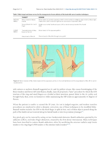
Figure 2. Nerve transfer of the motor branch of the opponens pollicis to the terminal division of the deep branch of the UN for pinch
reconstruction.
with sutures or anchors; Bunnell suggested an A1 and A2 pulleys release, this causes bowstringing of the
flexor tendons and favors MP joint flexion; finally, Zancolli proposed a “lasso” procedure by which the FDS
tendons of the ring and small fingers are divided at their insertion, passed distal to the A1 pulley and
brought back, then, sewn on themselves while maintaining the MP joints in approximately 60 degrees of
flexion.
When the patient is unable to extend the IP joint, the test is judged negative, and tendon transfers
procedures are employed to allow a dynamic correction; one of these techniques is the modified Stiles–
Bunnell tendon transfer: the FDS to the third finger is split in four, each of these slips is passed along the
[55]
path of the lumbrical and inserted along the lateral bands or into the proximal phalanges .
Key pinch grip can be restored by acting on two fundamental elements: thumb adduction, granted by the
adductor pollicis, and index finger abduction, ensured by the first dorsal interosseous. Many techniques
have been described to restore thumb adduction, either by sacrificing the extensor radialis carpi brevis
tendon or the ring finger’s FDS tendon or the extensor indicis tendon [58-60] .