
Page 67 - Read Online
P. 67
Titolo et al. Plast Aesthet Res 2023;10:21 https://dx.doi.org/10.20517/2347-9264.2022.113 Page 5 of 13
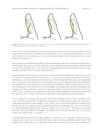
Figure 1. Digital neuroma following sharp-force trauma; neuroma has been excised, trimming the proximal and distal stumps, then an
end-to-end tension-free neurorrhaphy has been performed.
donor sites for these procedures are the sural, saphenous, sensory branch of the RN, medial or lateral
antebrachial cutaneous nerves, and the posterior interosseous nerve; in particular, the last two nerves are the
most suitable in the reconstruction of common and proper digital nerves because their loss is poorly
relevant for the patient [24,33-35] .
Despite being the gold standard procedure, several disadvantages have been reported in autologous nerve
grafts employ, such as scarcity of donor material, increased surgical time for harvesting the autograft, graft
and donor-site morbidity, loss of nerve function and potential neuroma formation in the donor site, and
possible mismatch between donor and recipient nerves [26,27,36] .
Vascularized nerve grafts in nerve reconstruction are employed in brachial plexus disorders, and their use in
the distal wrist and hand districts is uncommon. As a result, new solutions have been developed as
alternatives to autologous graft; commercial options consist in bridging the defect through biodegradable
nerve conduits composed of polymer-based materials or processed decellularized human nerve allografts.
Biodegradable nerve conduits protect the growing axons through a semipermeable cavity and create a
microenvironment rich in chemical factors that upregulate nerve regeneration; decellularized human nerve
allografts provide an internal microstructure and extracellular matrix molecules of the native nerve
tissue . Nerve conduits are easily applied in the nerve gaps by positioning sutures through the conduit and
[37]
the epineurium in a “U” fashion, whereas nerve allografts are placed similarly to autologous grafts .
[24]
Clinical outcomes on collagen conduits and processed nerve allografts have been widely released over the
years; even if these studies often use different scoring systems in their assessment that may render direct
confrontation problematic with variable recovery rates, it has been shown that globally all studies report
relevant recovery. For collagen conduits, excellent to good recovery rates ranges from 50% to 89%, while for
processed decellularized human nerve allografts, it spans from 83% to 100% [36-38] . Although good outcomes
have been associated with these techniques, their application is currently limited to non-critical, small
caliber, nerve defects repair (< 3 cm gaps) [26,32,36,37] .
A valuable solution for non-critical nerve gaps is the “muscle-in-vein” technique, which consists of an
[39]
autogenous vein graft used as a conduit filled with a stripe of autologous skeletal muscle tissue . The
benefits of this technique lie in the composition of both vein and muscle tissue: vein walls act perfectly as a