
Page 49 - Read Online
P. 49
Friedman et al. Plast Aesthet Res 2023;10:23 https://dx.doi.org/10.20517/2347-9264.2022.100 Page 7 of 15
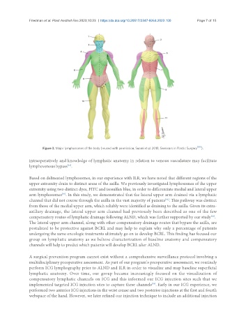
Figure 3. Major lymphosomes of the body (reused with permission, Suami et al. 2018, Seminars in Plastic Surgery [90] ).
intraoperatively and knowledge of lymphatic anatomy in relation to venous vasculature may facilitate
lymphovenous bypass .
[91]
Based on delineated lymphosomes, in our experience with ILR, we have noted that different regions of the
upper extremity drain to distinct areas of the axilla. We previously investigated lymphosomes of the upper
extremity using two distinct dyes, FITC and isosulfan blue, in order to differentiate medial and lateral upper
arm lymphosomes . In this study, we demonstrated that the lateral upper arm drained via a lymphatic
[92]
[92]
channel that did not course through the axilla in the vast majority of patients . This pathway was distinct
from those of the medial upper arm, which reliably were identified as draining to the axilla. Given its extra-
axillary drainage, the lateral upper arm channel had previously been described as one of the few
compensatory routes of lymphatic drainage following ALND, which was further supported by our study .
[92]
The lateral upper arm channel, along with other compensatory drainage routes that bypass the axilla, are
postulated to be protective against BCRL and may help to explain why only a percentage of patients
undergoing the same oncologic treatments ultimately go on to develop BCRL. This finding has focused our
group on lymphatic anatomy as we believe characterization of baseline anatomy and compensatory
channels will help to predict which patients will develop BCRL after ALND.
A surgical prevention program cannot exist without a comprehensive surveillance protocol involving a
multidisciplinary preoperative assessment. As part of our program’s preoperative assessment, we routinely
perform ICG lymphography prior to ALND and ILR in order to visualize and map baseline superficial
lymphatic anatomy. Over time, our group became increasingly focused on the visualization of
compensatory lymphatic channels on ICG and this informed our ICG injection sites such that we
[93]
implemented targeted ICG injection sites to capture these channels . Early in our ICG experience, we
performed two anterior ICG injections in the wrist crease and two posterior injections at the first and fourth
webspace of the hand. However, we later refined our injection technique to include an additional injection