
Page 60 - Read Online
P. 60
Page 56 Al-Sammarraie et al. Neuroimmunol Neuroinflammation 2021;8:53-63 I http://dx.doi.org/10.20517/2347-8659.2020.34
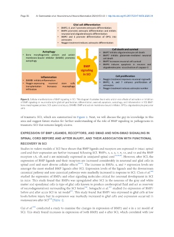
Figure 2. Cellular manifestations of BMP signaling in SCI. This diagram illustrates the in vitro and in vivo effects of activation or inhibition
of BMP signaling on neuronal and/or glial cell proliferation, differentiation, survival, apoptosis, autophagy, and inflammation in SCI. BMP:
bone morphogenic protein; SCI: spinal cord injury; BAMBI: BMP and activin membrane-bound inhibitor; OPCs: oligodendrocyte precursor
cells
of traumatic SCI, which are summarized in Figure 2. Next, we will discuss the gap in knowledge in this
area and suggest future studies for further understanding of the role of BMP signaling in pathogenesis in
traumatic SCI that remains largely elusive.
EXPRESSION OF BMP LIGANDS, RECEPTORS, AND SMAD AND NON-SMAD SIGNALING IN
SPINAL CORD BEFORE AND AFTER INJURY, AND THEIR ASSOCIATION WITH FUNCTIONAL
RECOVERY IN SCI
Studies in rodent models of SCI have shown that BMP ligands and receptors are expressed in intact spinal
cord and their expression are further increased following SCI. BMP2, 3, 4, 5, 7, 9, 12, and 13 and the BMP
receptors 1A, 1B, and 2 are minimally expressed in uninjured spinal cord [16,23,26] . However, after SCI, the
expression of BMP ligands and their receptors are increased considerably in neuronal and glial cells in
spinal cord and exert diverse cellular effects [23,27] . The increase in BMP2, 4, and 7 expression levels are
amongst the most studied BMP ligands after SCI. Expression levels of the ligands and the downstream
[28]
canonical pathway and non-canonical pathways were markedly increased in response to SCI. Chen et al.
studied the expression of BMP4 and other signaling molecules critical for neuronal development in SCI
in mice. This study found that BMP4 was upregulated after SCI in the neurons of the gray and white
matter and ependymal cells (a type of glial cells known to produce cerebrospinal fluid and act as reservoir
[27]
[28]
of neurodegeneration) surrounding the SCI lesion . Setoguchi et al. studied the expression of BMP7
[27]
before and after acute SCI in rat model . This study found that BMP7 was expressed in glial cells at low
levels before injury but its expression was markedly increased in glial cells and expression occurred in
[27]
motoneurons after SCI [Table 1].
[29]
Cui et al. conducted a study to examine the changes in expression of BMP2 and 4 in a rat model of
SCI. This study found increases in expression of both BMP2 and 4 after SCI, which correlated with low