
Page 8 - Read Online
P. 8
Davis et al. Neuroimmunol Neuroinflammation 2020;7:300-10 I http://dx.doi.org/10.20517/2347-8659.2020.19 Page 303
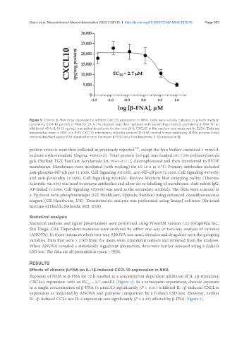
Figure 1. Chronic β-FNA dose-dependently inhibits CXCL10 expression in NHA. Cells were initially cultured in growth medium
containing 0.04-10 µmol/L β-FNA for 24 h; the medium was then replaced with serum-free medium containing β-FNA for an
additional 48 h. IL-1β (3 ng/mL) was added to cultures for the final 24 h. CXCL10 in the medium was measured by ELISA. Data are
presented as mean ± SEM (n = 8-9). CXCL10: interferon-γ inducible protein-10; NHA: normal human astrocytes; ELISA: enzyme-linked
immunoabsorbant assay; SEM: standard error of the mean; β-FNA: beta-funaltrexamine; IL-1β: interleukin-1β
[33]
protein extracts were then collected as previously reported , except the lysis buffers contained 1 mmol/L
sodium orthovanadate (Sigma, #450243). Total protein (30 µg) was loaded on 7.5% polyacrylamide
gels (BioRad TGX FastCast Acrylamide kit, #161-0171), electrophoresed and then transferred to PVDF
membranes. Membranes were incubated (with rocking) for 15-18 h at 4 °C. Primary antibodies included
anti-phospho-NF-κB p65 (1:1000, Cell Signaling #3033S), anti-NF-κB p65 (1:1000, Cell Signaling #4764S),
and anti-β-tubulin (1:1000, Cell Signaling #2146S). Restore Western blot stripping buffer (Thermo
Scientific #21059) was used to remove antibodies and allow for re-labelling of membranes. Anti-rabbit IgG,
AP-linked (1:1000, Cell Signaling #7054S) was used as the secondary antibody. The blots were scanned in
a Typhoon 9410 phosphorimager (GE Healthcare, Uppsala, Sweden) using enhanced chemifluorescence
reagent (GE Healthcare, UK). Densitometric analysis was performed using ImageJ software (National
Institute of Health, Bethesda, MD, USA).
Statistical analysis
Statistical analyses and figure presentations were performed using PrismTM version 7.04 (GraphPad Inc.,
San Diego, CA). Dependent measures were analyzed by either one-way or two-way analysis of variance
(ANOVA). In those instances where two-way ANOVA was used, stimulus and drug dose were the grouping
variables. Data that were > 2 SD from the mean were considered outliers and removed from the analyses.
When ANOVA revealed a statistically significant interaction, data were further assessed using a Fisher’s
LSD test. The data are all presented as mean ± SEM.
RESULTS
Effects of chronic β-FNA on IL-1β-induced CXCL10 expression in NHA
Exposure of NHA to β-FNA for 72 h resulted in a concentration-dependent inhibition of IL-1β-stimulated
CXCL10 expression, with an EC = 2.7 µmol/L [Figure 1]. In a subsequent experiment, chronic exposure
50
to a single concentration of β-FNA (3 µmol/L) significantly (P < 0.01) inhibited IL-1β-induced CXCL10
expression as indicated by ANOVA and pairwise comparison by a Fisher’s LSD test. However, neither
IL-1β-induced CCL2 nor IL-6 expression was significantly (P ≥ 0.22) affected by β-FNA [Figure 2].