
Page 11 - Read Online
P. 11
Page 306 Davis et al. Neuroimmunol Neuroinflammation 2020;7:300-10 I http://dx.doi.org/10.20517/2347-8659.2020.19
P
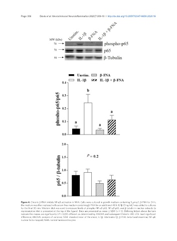
Figure 4. Chronic β-FNA inhibits NF-κB activation in NHA. Cells were cultured in growth medium containing 3 µmo/L β-FNA for 24 h;
the medium was then replaced with serum-free medium containing β-FNA for an additional 48 h. IL-1β (3 ng/mL) was added to cultures
for the final 30 min. Western blot was used to measure levels of phospho-NF-κB p65, NF-κB p65, and β-tubulin in nuclear extracts (a
representative blot is presented at the top of the figure). Data are presented as mean ± SEM (n = 5). Differing letters above the bars
indicate the means are significantly (P < 0.05) different as determined by ANOVA and subsequent Fisher’s LSD. LDS: least significant
difference; ANOVA: analysis of variance; SEM: standard error of the mean; IL-1β: interleukin-1β; β-FNA: beta-funaltrexamine; NF-κB:
nuclear factor-kappaB; NHA: normal human astrocytes