
Page 7 - Read Online
P. 7
Fracaro et al. Neuroimmunol Neuroinflammation 2020;7:1-12 I http://dx.doi.org/10.20517/2347-8659.2019.009 Page 3
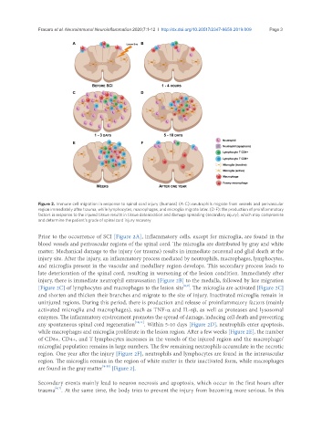
Figure 2. Immune cell migration in response to spinal cord injury (humans) (A-C): neutrophils migrate from vessels and perivascular
region immediately after trauma, while lymphocytes, macrophages, and microglia migrate later. (D-F): the production of proinflammatory
factors in response to the injured tissue results in tissue deterioration and damage spreading (secondary injury), which may compromise
and determine the patient’s grade of spinal cord injury recovery
Prior to the occurrence of SCI [Figure 2A], inflammatory cells, except for microglia, are found in the
blood vessels and perivascular regions of the spinal cord. The microglia are distributed by gray and white
matter. Mechanical damage to the injury (or trauma) results in immediate neuronal and glial death at the
injury site. After the injury, an inflammatory process mediated by neutrophils, macrophages, lymphocytes,
and microglia present in the vascular and medullary region develops. This secondary process leads to
late deterioration of the spinal cord, resulting in worsening of the lesion condition. Immediately after
injury, there is immediate neutrophil extravasation [Figure 2B] to the medulla, followed by late migration
[8,9]
[Figure 2C] of lymphocytes and macrophages to the lesion site . The microglia are activated [Figure 2C]
and shorten and thicken their branches and migrate to the site of injury. Inactivated microglia remain in
uninjured regions. During this period, there is production and release of proinflammatory factors (mainly
activated microglia and macrophages), such as TNF-α and IL-6β, as well as proteases and lysosomal
enzymes. The inflammatory environment promotes the spread of damage, inducing cell death and preventing
any spontaneous spinal cord regeneration [10,11] . Within 5-10 days [Figure 2D], neutrophils enter apoptosis,
while macrophages and microglia proliferate in the lesion region. After a few weeks [Figure 2E], the number
of CD8+, CD4+, and T lymphocytes increases in the vessels of the injured region and the macrophage/
microglial population remains in large numbers. The few remaining neutrophils accumulate in the necrotic
region. One year after the injury [Figure 2F], neutrophils and lymphocytes are found in the intravascular
region. The microglia remain in the region of white matter in their inactivated form, while macrophages
are found in the gray matter [8-11] [Figure 2].
Secondary events mainly lead to neuron necrosis and apoptosis, which occur in the first hours after
[6,7]
trauma . At the same time, the body tries to prevent the injury from becoming more serious. In this