
Page 35 - Read Online
P. 35
Benusa et al. Neuroimmunol Neuroinflammation 2020;7:23-39 I http://dx.doi.org/10.20517/2347-8659.2019.28 Page 31
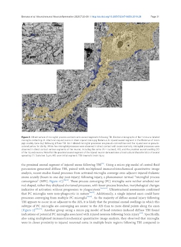
Figure 2. Ultrastructure of microglial process contacts onto axonal segments following TBI. Electron micrographs of Iba-1 immuno-labeled
microglia contacting: A: intact non-injured axons in sham-injured micro pig thalamus; B: injured axonal segment in the thalamus of micro
pigs acutely (one day) following diffuse TBI. Iba-1-labeled microglial processes are pseudo-colored blue and the injured axon is pseudo-
colored yellow for clarity. While few microglial processes were observed in direct contact with axons normally, microglial processes were
observed in direct contact various segments of the neuron, including the soma (N = nucleus), AIS, and the proximal axonal swelling (X)
of the injured neuron. Note that the proximal axonal segment of the injured neuron demonstrates ultrastructural characteristics of axonal
sprouting (*). Scale bar: 5 μm. AIS: axon initial segment; TBI: traumatic brain injury
[51]
the proximal axonal segment of injured axons following TBI . Using a micro pig model of central fluid
percussion-generated diffuse TBI, paired with multiplexed immunohistochemical quantitative image
analysis, recent studies found processes from activated microglia converge onto adjacent injured thalamic
axons acutely (hours to one day post-injury) following injury, a phenomenon termed “microglial process
convergence” (MPC; Figure 1C) [50,52] . These process converging (PC) microglia were neither ameboid nor
rod shaped, rather they displayed shortened processes, with fewer process branches, morphological changes
indicative of activation without progression to phagocytosis [13,51,52] . Ultrastructural assessments confirmed
that PC microglia were non-phagocytic in nature [50,52] . Additionally, a single injured axon could have
processes converging from multiple PC microglia [50,52] . As the majority of diffuse axonal injury following
TBI appears to occur in or adjacent to the AIS, it is likely that the proximal axonal swellings in which this
subtype of PC microglia are converging are nearer to the AIS than to more distal points along the axon
[Figure 2] [170,171] . Another group using a micro pig model of head-rotation-induced diffuse TBI found
[172]
indications of potential PC microglia associated with injured neurons following brain injury . Specifically,
also using multiplexed immunohistochemical quantitative image analysis, they observed that microglia
were in closer proximity to injured neuronal soma in multiple brain regions following TBI compared to