
Page 151 - Read Online
P. 151
Page 4 of 14 Zoghi et al. Neuroimmunol Neuroinflammation 2019;6:14 I http://dx.doi.org/10.20517/2347-8659.2019.03
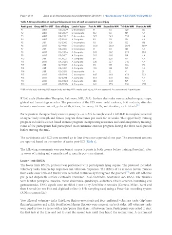
Table 2. Group allocation of each participant and time of each assessment post injury
Participant Group WBT or UBT Date of injury Level of injury First Ax WPI Second Ax WPI Third Ax WPI Fourth Ax WPI
P1 WBT 06/2009 D Incomplete 91 107 120 147
P2 WBT 03/2009 B Incomplete 152 167 NA NA
P3 WBT 04/2002 D Incomplete 527 543 553 NA
P4 WBT 07/2010 A Complete 83 113 124 NA
P5 UBT 10/2009 D Incomplete 93 108 122 148
P6 WBT 12/1960 D Incomplete 2641 2657 2673 2697
P7 UBT 08/2010 D Incomplete 91 107 118 NA
P8 WBT 06/2006 A Complete 243 262 273 300
P9 WBT 05/2001 A Complete 592 605 NA NA
P10 UBT 11/2002 A Complete 511 525 NA NA
P11 WBT 04/2006 A Complete 320 337 346 NA
P12 UBT 12/2008 A Complete 115 133 146 172
P13 UBT 08/2010 A Complete 109 122 NA NA
P14 UBT 06/2011 B Incomplete 6 22 41 61
P15 WBT 03/1999 C Incomplete 647 663 678 703
P16 WBT 10/2001 A Complete 554 570 580 NA
P17 WBT 08/2003 A Complete 418 433 NA NA
P18 WBT 09/1992/ A Complete 961 979 991 1019
WBT: whole body training; UBT: upper body training; WPI: weeks post injury; NA: not assessed; Ax: assessment; P: participant
RT300 cycle (Restorative Therapies, Baltimore, MD, USA). Surface electrodes were attached on quadriceps,
gluteal and hamstrings muscles. The parameters of the FES were: pedal cadence, 5-50 rev/min; stimulus
[24]
intensity, maximum 140 mA; pulse width, 0.3 ms; frequency, 35 Hz; and duration, up to 30 min .
Participants in the upper body exercise group (n = 6, 3 AIS A complete and 3 AIS B-D incomplete) received
an upper body strength and fitness program three times per week for 12 weeks. This upper body training
program included a circuit-based exercise program incorporating resistance and cardiorespiratory training.
None of the participants had participated in an intensive exercise program during the three-week period
before starting this trial.
The participants with SCI were assessed up to four times over a period of one year. The assessment sessions
are reported based on the number of weeks post-SCI [Table 2].
The following assessments were performed on participants in both groups before training (baseline), after
12 weeks of training and 6 months and 12 months post-recruitment.
Lower limb BMCA
The lower limb BMCA protocol was performed with participants lying supine. The protocol included:
voluntary tasks, tendon-tap responses and vibration responses. The sEMG of 14 muscles (seven muscles
[21]
from each lower limb and trunk) were recorded continuously throughout the protocol with self-adhesive
pre-gelled disposable surface electrodes (Noraxon Dual electrodes, Scottsdale AZ, USA). The muscles
were lumbar paraspinal muscles, rectus abdominis, quadriceps, adductors, tibialis anterior, hamstring and
gastrocnemius. EMG signals were amplified (1000 ×) by ZeroWire electrodes (Cometa, Milan, Italy) and
then filtered (20-500 Hz) and digitised online (1 kHz sampling rate) using a PowerLab recording system
(ADInstruments Ltd).
Two bilateral voluntary tasks (hip/knee flexion-extension) and four unilateral voluntary tasks (hip/knee
flexion/extension and ankle dorsiflexion/plantar flexion) were assessed on both sides. All voluntary tasks
were cued by two 5-s tones with a brief pause (less than 1 s) between them. Participants were asked to start
the first task at the tone and not to start the second task until they heard the second tone. A customised