
Page 139 - Read Online
P. 139
Alsulihem et al. Neuroimmunol Neuroinflammation 2019;6:13 I http://dx.doi.org/10.20517/2347-8659.2019.007 Page 5 of 13
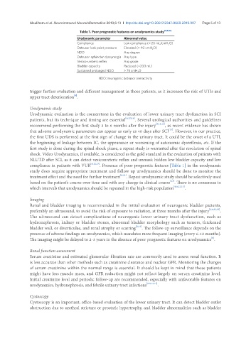
Table 1. Poor prognostic features on urodynamics study [6,8,12]
Urodynamic parameter Abnormal value
Compliance Low compliance (< 20 mL/cmH 2 O)
Detrusor leak point pressure Elevated (> 40 cmH 2 O)
NDO Any degree
Detrusor-sphincter dyssynergia Any type
Vesico-ureteric reflex Any grade
Bladder capacity Reduced (<200 mL)
Sustained prolonged NDO > 75 cmH 2 O
NDO: neurogenic detrusor overactivity
trigger further evaluation and different management in those patients, as it increases the risk of UTIs and
[8]
upper tract deterioration .
Urodynamic study
Urodynamic evaluation is the cornerstone in the evaluation of lower urinary tract dysfunction in SCI
patients, but its technique and timing are essential [6,12,14] . Several urological authorities and guidelines
recommend performing the first study 3 to 6 months after the injury [8,12,14] , as recent evidence has shown
that adverse urodynamic parameters can appear as early as 40 days after SCI . However, in our practice,
[15]
the first UDS is performed at the first sign of change in the urinary tract. It could be the onset of a UTI,
the beginning of leakage between IC, the appearance or worsening of autonomic dysreflexia, etc. If the
first study is done during the spinal shock phase, a repeat study is warranted after the resolution of spinal
shock. Video Urodynamics, if available, is considered as the gold standard in the evaluation of patients with
NLUTD after SCI, as it can detect vesicoureteric reflux and unmask hidden low bladder capacity and low
compliance in patients with VUR [6,12,13] . Presence of poor prognostic features [Table 1] in the urodynamic
study does require appropriate treatment and follow up urodynamics should be done to monitor the
treatment effect and the need for further treatment [6,12] . Repeat urodynamic study should be selectively used
[13]
based on the patient’s course over time and with any change in clinical course . There is no consensus in
which intervals that urodynamics should be repeated in the high-risk population [6,8,12,13] .
Imaging
Renal and bladder imaging is recommended in the initial evaluation of neurogenic bladder patients,
preferably an ultrasound, to avoid the risk of exposure to radiation, at three months after the injury [6,8,12,13] .
The ultrasound can detect complications of neurogenic lower urinary tract dysfunction, such as
hydronephrosis, kidney or bladder stones, abnormal bladder morphology such as tumors, thickened
bladder wall, or diverticulae, and renal atrophy or scarring [8,12] . The follow-up surveillance depends on the
presence of adverse findings on urodynamics, which mandates more frequent imaging (every 6-12 months).
[6]
The imaging might be delayed to 2-3 years in the absence of poor prognostic features on urodynamics .
Renal function assessment
Serum creatinine and estimated glomerular filtration rate are commonly used to assess renal function. It
is less accurate than other methods such as creatinine clearance and nuclear GFR. Monitoring the changes
of serum creatinine within the normal range is essential. It should be kept in mind that those patients
might have less muscle mass, and GFR reduction might not reflect largely on serum creatinine level.
Initial creatinine level and periodic follow-up are recommended, especially with unfavorable features on
urodynamics, hydronephrosis, and febrile urinary tract infections [6,8,12,13] .
Cystoscopy
Cystoscopy is an important, office-based evaluation of the lower urinary tract. It can detect bladder outlet
obstruction due to urethral stricture or prostatic hypertrophy, and bladder abnormalities such as bladder