
Page 129 - Read Online
P. 129
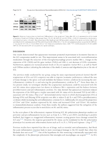
Page 8 of 13               Souza et al. Neuroimmunol Neuroinflammation 2019;6:12  I  http://dx.doi.org/10.20517/2347-8659.2019.04

                A                                 B                            C

                                     BV
                       Sham        CTL     BV (NP)  (ST36+GV3)


               Figure 5. Influence of apipuncture on NeuN and CNPase levels in the spinal cord 7 days after SCI. In A: representative of the bands
               of proteins of NeuN and CNPase by the groups Sham, CTL-SCI, BV (NP)-SCI and BV (ST36 + GV3)-SCI measured by densitometry,
               normalized by β-actin; In B and C: the comparison of the NeuN and anti-CNPase protein content at the spinal cord lesion site of rats
               submitted to to SCI and apipuncture at ST36 and GV3 points [BV (ST36)-SCI; n = 6], SCI and apipuncture at non-acupoints [BV (NP)-SCI,
               n = 6], only SCI (CTL-SCI, n = 6) and Sham-SCI (n = 6). Values are presented as mean ± SEM. ***P < 0.001 compared to CTL-SCI and BV
               (NP)-SCI. BV: bee venom; NP: non-acupoints; SCI: spinal cord injury


               DISCUSSION
               Our results demonstrated that apipuncture treatment promoted improvement in locomotor function in
               the SCI compression model in rats. This improvement seems to be associated with neuroinflammation
               modulation through the reduction of the microglia/macrophage protein marker IBA-1, changes in the
               expression of M1 (iNOS) and M2 gene markers (TGF-β and ARG-1), and decrease of COX-2 expression.
               Furthermore, apipuncture increased protein levels of the anti-apoptotic marker BCL-2, as well as NeuN
               and CNPase markers indicating the reduction of the death of neurons and oligodendrocytes at the site of
               SCI.

               The previous study conducted by our group, using the same experimental protocol showed that BV
               acupuncture at ST36 and GV3 acupoints was able to improve locomotor performance, reduced the area
               of tissue damage in the spinal cord and modulate the balance between cytokines by increasing the anti-
               inflammatory cytokine IL-10 and reducing the proinflammatory cytokine IL-6 . From these data, in
                                                                                     [17]
               the present study, we investigate whether the acupuncture mechanism could be associated with M1
               and M2 status since apipuncture has shown to influence IBA-1 expression and the balance between
               proinflammatory and anti-inflammatory cytokine. Our data showed that apipuncture treatment reduced
               the expression of the iNOS gene a marker associated with M1 status whilst decreased TGF-β and ARG-1,
                                                [7]
               associated with M2 status. Zhao et al.  demonstrated similar results in which electroacupuncture (EA) at
               Jizhong (GV6) and Zhiyang (GV9) acupoints was able to reduce IL-1β, IL-6 and TNF-α proinflammatory
               cytokines whilst increasing the anti-inflammatory cytokine IL-10. Additionally, EA reduced the proportion
                                                                                              +
                                                                                  +
                                 +
                       +
               of CD68  and CD86  markers expressed by M1 status and increased CD68  and CD206  M2 markers
               in immunohistochemical analysis. From these results, the authors suggested that the mitigation of the
               inflammatory process occurs due to modulation on M1 and M2 status.

               In the acute phase of the inflammatory response following SCI, M1 polarization phenotype markers are
               prevalent and pro-inflammatory factors such as IL1β, IL-6, TNF-α and iNOS contributing to positive
               feedback that triggers an exaggerated inflammatory response causing greater tissue damage around the
               SCI area [22,25] . However, the response generated by M1 status can be modulated by the M2 polarization
               phenotype that secretes anti-inflammatory factors such as IL-10, IL13, TGF-β and Arginase-1 (Arg-1) [18,27,34] .


               Previous studies have reported that the use of drugs, such as steroidal anti-inflammatory, that block
               the acute inflammatory response leads to more neurological damage in the SCI model than when this
               blockade does not occur [18,20,35] . One of the hypotheses for this worsening is intrinsically associated with
               the complexity of the phenotypic polarization that microglia and macrophages can acquire [18,21,25] . In SCI,