
Page 304 - Read Online
P. 304
Chowdhury et al. Neuroimmunol Neuroinflammation 2018;5:39 I http://dx.doi.org/10.20517/2347-8659.2018.37 Page 3 of 5
A A B
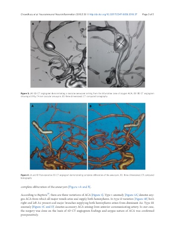
Figure 3. (A) 3D CT angiogram demonstrating a saccular aneurysm arising from the bifurcation zone of azygos ACA; (B) 3D CT angiogram
showing a 5.9 by 7.4 mm saccular aneurysm. 3D: three dimensional; CT: computed tomography
A B
Figure 4. (A and B) Post-operative 3D CT angiogram demonstrating complete obliteration of the aneurysm. 3D: three dimensional; CT: computed
tomography
complete obliteration of the aneurysm [Figure 4A and B].
[7]
According to Baptista , there are three variations of ACA [Figure 5]. Type 1 anomaly [Figure 5A] denotes azy-
gos ACA from which all major vessels arise and supply both hemispheres. In type II variation [Figure 5B] both
right and left A2 present and major branches supplying both hemispheres arises from dominant A2. Type III
anomaly [Figure 5C and D] denotes accessory ACA arising from anterior communicating artery. In our case,
the surgery was done on the basis of 3D CT angiogram findings and azygos nature of ACA was confirmed
preoperatively.