
Page 303 - Read Online
P. 303
Page 2 of 5 Chowdhury et al. Neuroimmunol Neuroinflammation 2018;5:39 I http://dx.doi.org/10.20517/2347-8659.2018.37
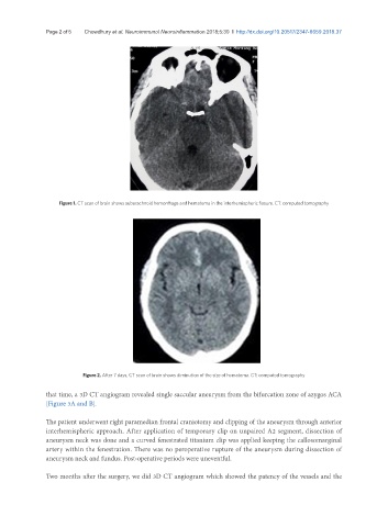
Figure 1. CT scan of brain shows subarachnoid hemorrhage and hematoma in the interhemispheric fissure. CT: computed tomography
Figure 2. After 7 days, CT scan of brain shows diminution of the size of hematoma. CT: computed tomography
that time, a 3D CT angiogram revealed single saccular aneurysm from the bifurcation zone of azygos ACA
[Figure 3A and B].
The patient underwent right paramedian frontal craniotomy and clipping of the aneurysm through anterior
interhemispheric approach. After application of temporary clip on unpaired A2 segment, dissection of
aneurysm neck was done and a curved fenestrated titanium clip was applied keeping the callosomarginal
artery within the fenestration. There was no peroperative rupture of the aneurysm during dissection of
aneurysm neck and fundus. Post-operative periods were uneventful.
Two months after the surgery, we did 3D CT angiogram which showed the patency of the vessels and the