
Page 227 - Read Online
P. 227
Page 4 of 7 Chen et al. Neuroimmunol Neuroinflammation 2018;5:31 I http://dx.doi.org/10.20517/2347-8659.2018.23
MRI-T2
MRI-DWI
MRI-SWI
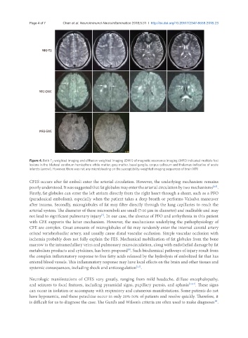
Figure 4. Both T 2 -weighted imaging and diffusion weighted imaging (DWI) of magnetic resonance imaging (MRI) indicated multiple foci
lesions in the bilateral cerebrum hemisphere white matter, grey matter, basal ganglia, corpus callosum and thalamus indicative of acute
infarcts (arrow). However, there was not any microbleeding on the susceptibility-weighted imaging sequences of brain MRI
CFES occurs after fat emboli enter the arterial circulation. However, the underlying mechanism remains
poorly understood. It was suggested that fat globules may enter the arterial circulation by two mechanisms .
[2,7]
Firstly, fat globules can enter the left atrium directly from the right heart through a shunt, such as a PFO
(paradoxical embolism), especially when the patient takes a deep breath or performs Valsalva maneuver
after trauma. Secondly, microglobules of fat may filter directly through the lung capillaries to reach the
arterial system. The diameter of these microemboli are small (7-10 μm in diameter) and malleable and may
not lead to significant pulmonary injury . In our case, the absence of PFO and arrhythmia in this patient
[7]
with CFE supports the latter mechanism. However, the mechanisms underlying the pathophysiology of
CFE are complex. Great amounts of microglobules of fat may randomly enter the internal carotid artery
or/and vertebrobasilar artery, and usually cause distal vascular occlusion. Simple vascular occlusion with
ischemia probably does not fully explain the FES. Mechanical mobilization of fat globules from the bone
marrow to the intramedullary veins and pulmonary microcirculation, along with endothelial damage by fat
metabolism products and cytokines, has been proposed . Such biochemical pathways of injury result from
[8]
the complex inflammatory response to free fatty acids released by the hydrolysis of embolized fat that has
entered blood vessels. This inflammatory response may have local effects on the brain and other tissues and
systemic consequences, including shock and anticoagulation .
[2,7]
Neurologic manifestations of CFES vary greatly, ranging from mild headache, diffuse encephalopathy,
and seizures to focal features, including pyramidal signs, pupillary paresis, and aphasia [1,2,7] . These signs
can occur in isolation or accompany with respiratory and cutaneous manifestations. Some patients do not
have hypoxemia, and these petechiae occur in only 20%-50% of patients and resolve quickly. Therefore, it
is difficult for us to diagnose the case. The Gurd's and Wilson's criteria are often used to make diagnoses .
[9]