
Page 184 - Read Online
P. 184
Hirschberg et al. Neuroimmunol Neuroinflammation 2018;5:27 I http://dx.doi.org/10.20517/2347-8659.2018.31 Page 3 of 11
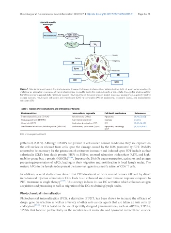
Figure 1. Mechanism and targets for photodynamic therapy. Following photosensitizer administration, light of a particular wavelength
matching an absorption resonance of the photosensitizer, is used to excite the molecule up to a triple state. The excited photosensitizer
3
1
transfers energy to ground state molecular oxygen ( O 2 ) resulting in the generation of singlet molecular oxygen ( O 2 ), a potent reactive
oxygen species, resulting in cell death. cell membrane (CM) mitochondria (Mito), endosome, lysosome (Lyso), and endoplasmic
reticulum (ER)
Table 1. Typical photosensitizers and intracellular targets
Photosensitizer Intra-cellular organelle Cell death mechanism References
5-aminolaevulinic acid (5-ALA) Mitochondria (Mito) Apoptosis [8,46,50,65]
Hematoporphyrin (HMME) Cell membrane (CM) necrosis [7,8,51]
Hypericin (HYP) Endoplasmic reticulum (ER) ICD [8,20,54,58]
Disulfonated aluminum phthalocyanine (AlPcS2a) Endosomes, Lysosomes (Lyso) Apoptosis, autophagy [8,15,29,31,62]
ICD?
ICD: immunogenic cell death
patterns (DAMPs). Although DAMPs are present in cells under normal conditions, they are exposed on
the cell surface or released from cells upon the damage caused by the ROS generated by PDT. DAMPs
reported to be necessary for the generation of antitumor immunity and induced upon PDT include surface
calreticulin (CRT), heat shock protein (HSP) 70, HSP90, secreted adenosine triphosphate (ATP), and high-
mobility group box 1 protein (HMGB1) [21-25] . Importantly, DAMPs cause maturation, activation and antigen
processing/presentation of APCs, leading to their migration and proliferation in local lymph nodes. The
+
mature APCs in the lymph nodes present the tumor antigens to a specific subset of CD8 T cells.
In addition, several studies have shown that PDT-treatment of extra cranial tumors followed by direct
intra-tumoral injection of immature DCs, leads to an enhanced anti-tumor immune response compared to
PDT treatment as single therapy [26-28] . This strategy induces in situ DC activation which enhances antigen
acquisition and processing as well as migration of the DCs to draining lymph nodes.
Photochemical internalization
Photochemical internalization (PCI), a derivative of PDT, has been shown to increase the efficacy of
drugs, gene transfection as well as a variety of other anti-cancer agents that are taken up into cells by
endocytosis [29-33] . PCI is based on the use of specially designed photosensitizers, such as AlPcS2a, TPPS2a,
TPcS2a that localize preferentially in the membranes of endocytic and lysosomal intracellular vesicles.