
Page 176 - Read Online
P. 176
Arora et al. Neuroimmunol Neuroinflammation 2018;5:26 I http://dx.doi.org/10.20517/2347-8659.2018.11 Page 3 of 8
Edge enhanced
Edge blurred
X
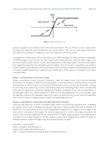
Figure 2. Image enhancement curve
proposes a graphical user interface (GUI) with interactive buttons. The use of GUI in such a system makes
the task of executing the whole classification very easy for anyone. The user can easily segment the tumor
and classify it to be benign or malignant in just clicks of buttons and within seconds.
An image from a MRI scanner is received in the form of a DICOM image. For better and faster computation,
a DICOM image is converted into the JPEG format before being processed. Then the JPEG image is pre-
processed mainly for the removal of noise and enhancement of the image quality. The processed image is
then segmented using the sub-pixel edge detector method. Then the required computations are made to
evaluate multiple values like mean, standard deviation, IDM, skewness, correlation, and homogeneity. Finally
a k-nearest neighbor algorithm is applied for the classification of the tumor in accordance to the above
computed values.
Stage 1: enhancement of the tumor image
Image enhancement is used in medical imaging to make the images clearer and to ensure optimum
presentation of all digital computer processing [Figure 2]. The importance of enhancement is to aid the
interpretation by both humans and computers. Enhancement aims at improving the quality of image
by removing noise, enhancing contrast, emphasizing edges and modifying shapes. Many computerized
techniques are widely used and applied including the histogram equalization, linear shift invariant filters and
morphological filters. For contrast enhancement there are two basic approaches, the first is Top-Hat, where
the algorithm enhances the segmented edges of the region of interest and the second where the algorithm
[7]
deals with the contrast of original image to enhance the segmentation process .
Stage 2: segmentation using sub-pixel edge detection technique
Sub-pixel edge detection consists of multiple stages within itself including edge detection, computing
the gradient vector field, computing the sub-pixel edge points, chaining edge points and thresholds with
[5]
hysteresis . The main motive of the use of an edge detection algorithm is to segment out the edges of the
tumor which is detected in the image. The sub-pixel edge detection works on the edges detected by the
Canny or the Devernay algorithm for the refinement of the edges using the region-based segmentation
techniques on the edges themselves. The line segments consisting of multiple pixels within the edges are
[8]
basically modified to depict the edges using the minimum number of pixels .
Algorithm 1: image_gradient
Input - an image I, scale parameter S;
Output - the image gradient field vector g;
Step 1 - compute and derive the Gaussian filter of standard deviation S;