
Page 175 - Read Online
P. 175
Page 2 of 8 Arora et al. Neuroimmunol Neuroinflammation 2018;5:26 I http://dx.doi.org/10.20517/2347-8659.2018.11
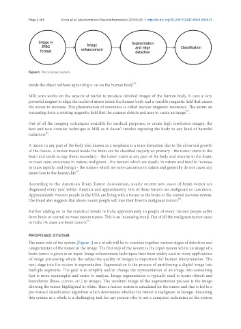
Image in Segmentation
JPEG Image and edge Classification
format enhancement detection
Figure 1. The proposed system
[2]
inside the object without appending a cut on the human body .
MRI scan works on the aspects of nuclei to produce detailed images of the human body. It uses a very
powerful magnet to align the nuclei of atoms inside the human body and a variable magnetic field that causes
the atoms to resonate. This phenomenon of resonance is called nuclear magnetic resonance. The atoms on
resonating form a rotating magnetic field that the scanner detects and uses to create an image .
[3]
Out of all the imaging techniques available for medical purposes, to create high resolution images, the
best and non-invasive technique is MRI as it doesn’t involve exposing the body to any kind of harmful
[2]
radiations .
A tumor in any part of the body also known as a neoplasm is a mass formation due to the abnormal growth
of the tissues. A tumor found inside the brain can be classified majorly as: primary - the tumor starts in the
brain and tends to stay there; secondary - the tumor starts at any part of the body and traverse to the brain,
in most cases cancerous in nature; malignant - the tumors which are deadly in nature and tend to increase
in mass rapidly; and benign - the tumors which are non-cancerous in nature and generally do not cause any
[4]
major loss to the human life .
According to the American Brain Tumor Association, nearly 80,000 new cases of brain tumor are
diagnosed every year within America and approximately 32% of these tumors are malignant or cancerous.
Approximately 700,000 people in the USA are living with a tumor in the brain or the central nervous system.
[5]
The trend also suggests that above 16,000 people will lose their lives to malignant tumors .
Further adding on to the statistical trends in India approximately 10 people of every 100,000 people suffer
from brain or central nervous system tumor. This is an increasing trend. Out of all the malignant tumor cases
[6]
in India 2% cases are brain tumors .
PROPOSED SYSTEM
The main role of the system [Figure 1] as a whole will be to combine together various stages of detection and
categorization of the tumor in the image. The first step of the system is the input system where an image of a
brain tumor is given as an input. Image enhancement techniques have been widely used in many applications
of image processing where the subjective quality of images is important for human interpretation. The
next stage into the system is segmentation. Segmentation is the process of partitioning a digital image into
multiple segments. The goal is to simplify and/or change the representation of an image into something
that is more meaningful and easier to analyze. Image segmentation is typically used to locate objects and
boundaries (lines, curves, etc.) in images. The resultant image of the segmentation process is the image
showing the tumor highlighted in white. Then a feature matrix is calculated for the tumor and that is fed to a
pre-trained classification algorithm which determines whether the tumor is malignant or benign. Executing
this system as a whole is a challenging task for any person who is not a computer technician so the system