
Page 206 - Read Online
P. 206
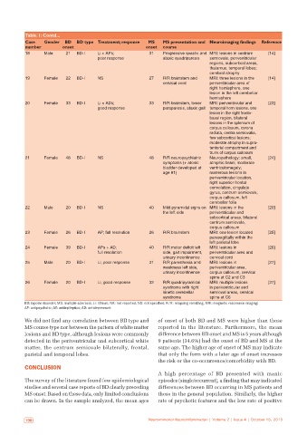
Table 1: Contd...
           Case   Gender  BD  BD type Treatment; response  MS   MS presentation and  Neuroimaging findings  Reference
           number        onset                            onset  course
           18     Male     21  BD-I    Li + APs;           31   Progressive spastic and  MRI: lesions in centrum   [14]
                                       poor response            ataxic quadriparesis  semiovale, periventricular
                                                                                  regions, subcortical areas,
                                                                                  thalamus, temporal lobes;
                                                                                  cerebral atrophy
           19     Female   22  BD-I    NS                  27   R/R brainstem and   MRI: three lesions in the   [14]
                                                                cervical cord     periventricular area of
                                                                                  right hemisphere, one
                                                                                  lesion in the left cerebellar
                                                                                  hemisphere
           20     Female   33  BD-I    Li + ADs;           33   R/R brainstem, lower   MRI: periventricular and   [23]
                                       good response            paraparesis, ataxic gait  temporal horn lesions, one
                                                                                  lesion in the right fronto-
                                                                                  basal region, bilateral
                                                                                  lesions in the splenium of
                                                                                  corpus callosum, corona
                                                                                  radiata, centra semiovalia,
                                                                                  few subcortical lesions;
                                                                                  moderate atrophy in supra-
                                                                                  tentorial compartment and
                                                                                  trunk of corpus callosum
           21     Female   48  BD-I    NS                  48   R/R neuropsychiatric   Neuropathology: small,   [24]
                                                                symptoms (+ atonic   atrophic brain; moderate
                                                                bladder developed at   ventriculomegaly;
                                                                age 81)           numerous lesions in
                                                                                  periventricular location,
                                                                                  right superior frontal
                                                                                  convolution, cingulate
                                                                                  gyrus, centrum semiovale,
                                                                                  corpus callosum, left
                                                                                  cerebellar folia
           22     Male     20  BD-I    NS                  40   Mild pyramidal signs on  MRI: lesions in the   [20]
                                                                the left side     periventricular and
                                                                                  subcortical areas, bilateral
                                                                                  centrum semiovale,
                                                                                  corpus callosum
           23     Female   26  BD-I    AP; full resolution  26  R/R brainstem     MRI: one lesion located   [25]
                                                                                  parasagittally within the
                                                                                  left parietal lobe
           24     Female   39  BD-I    APs + AD;           40   R/R motor deficit left   MRI: lesions in   [26]
                                       full resolution          side, gait impairment,   periventricular area and
                                                                urinary incontinence  cervical cord
           25     Male     20  BD-I    Li; poor response   31   R/R paresthesia and   MRI: lesions in    [27]
                                                                weakness left side,   periventricular area,
                                                                urinary incontinence  corpus callosum, cervical
                                                                                  spine at C2 and C3
           26     Female   20  BD-I    Li; good response   32   R/R quadripyramidal   MRI: multiple lesions   [27]
                                                                syndrome with right   in periventricular and
                                                                kinetic cerebellar   semioval areas, cervical
                                                                syndrome          spine at C6
           BD: bipolar disorder; MS: multiple sclerosis; Li: lithium; NR: not reported; NS: not specified; R/R: relapsing remitting; MRI: magnetic resonance imaging;
           AP: antipsychotic; AE: antiepileptics; AD: antidepressant

           We did not find any correlation between BD type and   of onset of both BD and MS were higher than those
           MS course type nor between the pattern of white matter   reported in the literature.  Furthermore,  the  mean
           lesions and BD type, although lesions were commonly   difference between BD onset and MS is 5 years although
           detected in the periventricular and subcortical white   9 patients (34.6%) had the onset of BD and MS at the
           matter, the centrum semiovale bilaterally, frontal,   same age. The higher age of onset of MS may indicate
           parietal and temporal lobes.                       that only the form with a later age of onset increases
                                                              the risk or the co‑occurrence/comorbidity with BD.
           CONCLUSION
                                                              A  high  percentage  of  BD  presented  with  manic
           The survey of the literature found few epidemiological   episodes (single/recurrent), a finding that may indicated
           studies and several case reports of BD clearly preceding   differences between BD occurring in MS patients and
           MS onset. Based on these data, only limited conclusions   those in the general population. Similarly, the higher
           can be drawn. In the sample analyzed, the mean ages   rate of psychotic features and the low rate of positive


            198                                           Neuroimmunol Neuroinflammation | Volume 2 | Issue 4 | October 15, 2015  Neuroimmunol Neuroinflammation | Volume 2 | Issue 4 | October 15, 2015                           199