
Page 205 - Read Online
P. 205
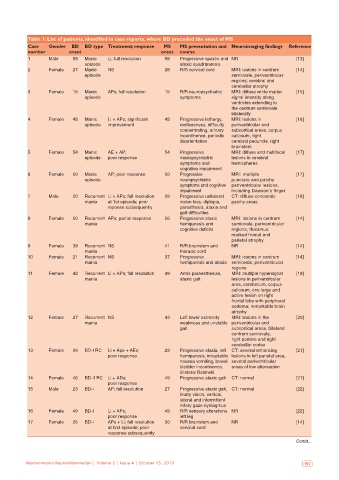
Table 1: List of patients, identified in case reports, where BD preceded the onset of MS
           Case   Gender  BD  BD type Treatment; response  MS   MS presentation and  Neuroimaging findings  Reference
           number        onset                            onset  course
           1      Male     58  Manic   Li; full resolution  58  Progressive spastic and  NR              [13]
                               episode                          ataxic quadriparesis
           2      Female   27  Manic   NS                  28   R/R cervical cord  MRI: lesions in centrum   [14]
                               episode                                            semiovale, periventricular
                                                                                  regions; cerebral and
                                                                                  cerebellar atrophy
           3      Female   15  Manic   APs; full resolution  15  R/R neuropsychiatric   MRI: diffuse white matter   [15]
                               episode                          symptoms          signal intensity along
                                                                                  ventricles extending to
                                                                                  the centrum semiovale
                                                                                  bilaterally
           4      Female   48  Manic   Li + APs; significant   48  Progressive lethargy,   MRI: lesions in   [16]
                               episode  improvement             restlessness, difficulty   periventricular and
                                                                concentrating, urinary   subcortical areas, corpus
                                                                incontinence, periodic   callosum, right
                                                                disorientation    cerebral peduncle, right
                                                                                  brainstem
           5      Female   54  Manic   AE + AP;            54   Progressive       MRI: diffuse and multifocal   [17]
                               episode  poor response           neuropsychiatric   lesions in cerebral
                                                                symptoms and      hemispheres
                                                                cognitive impairment
           6      Female   50  Manic   AP; poor response   50   Progressive       MRI: multiple          [17]
                               episode                          neuropsychiatric   punctate and patchy
                                                                symptoms and cognitive  periventricular lesions,
                                                                impairment        including Dawson’s finger
           7      Male     20  Recurrent  Li + APs; full resolution   29  Progressive unilateral   CT: diffuse concentric   [18]
                               mania   at 1st episode; poor     vision loss, diplopia,   patchy areas
                                       reponse subsequently     paresthesia, ataxia and
                                                                gait difficulties
           8      Female   50  Recurrent  APs; partial response  56  Progressive ataxic   MRI: lesions in centrum   [14]
                               mania                            hemiparesis and   semiovale, periventricular
                                                                cognitive deficits  regions, thalamus;
                                                                                  marked frontal and
                                                                                  parietal atrophy
           9      Female   39  Recurrent  NS               41   R/R brainstem and   NR                   [14]
                               mania                            thoracic cord
           10     Female   21  Recurrent  NS               37   Progressive       MRI: lesions in centrum   [14]
                               mania                            hemiparesis and ataxia  semiovale, periventricular
                                                                                  regions
           11     Female   42  Recurrent  Li + APs; full resolution  49  Arms paraesthesias,   MRI: multiple hypersignal   [19]
                               mania                            ataxic gait       lesions in periventricular
                                                                                  area, cerebellum, corpus
                                                                                  callosum, one large and
                                                                                  active lesion on right
                                                                                  frontal lobe with peripheral
                                                                                  oedema; remarkable brain
                                                                                  atrophy
           12     Female   27  Recurrent  NS               43   Left lower extremity   MRI: lesions in the   [20]
                               mania                            weakness and unstable  periventricular and
                                                                gait              subcortical areas, bilateral
                                                                                  centrum semiovale,
                                                                                  right pontine and right
                                                                                  cerebellar cortex
           13     Female   26  BD-I RC  Li + Aps + AEs;    29   Progressive ataxia, left  CT: several enhancing   [21]
                                       poor response            hemiparesis, intractable  lesions in left parietal area,
                                                                nausea vomiting, bowel  several periventricular
                                                                bladder incontinence,   areas of low attenuation
                                                                bilateral Babinski
           14     Female   46  BD-II RC  Li + ADs;         49   Progressive ataxic gait  CT: normal      [21]
                                       poor response
           15     Male     23  BD-I    AP; full resolution  27  Progressive ataxic gait,  CT: normal     [22]
                                                                blurry vision, vertical,
                                                                lateral and intermittent
                                                                rotary gaze nystagmus
           16     Female   49  BD-I    Li + APs;           49   R/R sensory alterations  NR              [22]
                                       poor response            left leg
           17     Female   25  BD-I    APs + Li; full resolution   30  R/R brainstem and   NR            [14]
                                       at first episode; poor   cervical cord
                                       response subsequently
                                                                                                         Contd...


   196  Neuroimmunol Neuroinflammation | Volume 2 | Issue 4 | October 15, 2015  Neuroimmunol Neuroinflammation | Volume 2 | Issue 4 | October 15, 2015   197