
Page 85 - Read Online
P. 85
were precursor events emerging 1-4 weeks before in muscle, deltoid, quadriceps, pretibial muscle, and
some of the patients: respiratory infection or fever gastrocnemius by observing whether there was a
in 126 cases (42.9%), digestive tract infection in spontaneous activity or not on the resting moment and
71 cases (24.1%), flu vaccination in 3 patients (1.0%), by testing the motor unit potential and recruitment
influenza vaccination in 2 cases (0.7%), rabies order of slight and strong muscle contraction. [2,3]
vaccination in 2 cases (0.7%), pregnancy in 1 case (0.3%),
and the rest cases with no precursor events. The first Diagnostic and evaluation criteria
symptom was limb weakness in 276 cases (93.9%), (1) GBS and nerve block were diagnosed under the
sensory disturbance in 98 cases (33.3%), and cranial diagnosis and treatment guidelines of GBS published in
nerve symptom in 54 cases (18.4%). There were 2010 by Chinese Medicine Association; (2) the neural
76 cases (25.9%) with clinical symptoms of cranial electro-physiological types were classified into AIDP
nerve lesions, including 36 cases with facial nerve and AMAN according to the electro-physiological
[4]
paralysis, 8 cases with ophthalmoplegia, 7 cases with diagnostic standard [Table 1]; (3) normal patients
diplopia and 62 cases with drinking water choking. and the patients not satisfying the diagnostic criteria
There were 102 cases (34.7%) with clinical symptoms of AIDP and AMAN were included into the unclear
of disturbance of sensation, 260 cases (88.4%) with type group; (4) the disease classification and follow-
upper-limb weakness, 276 cases (93.9%) with lower up results were marked according to rating scales
limb weakness. There were 192 cases (65.3%) with designed by Hughes et al. Based on patients’ ability to
tendon hyporeflexia, 66 cases (22.4%) with disappearing walk with the help, patients were classified into mild
and 11 cases (3.7%) with hyperreflexia. 261 cases had type and serious type. Patients with Hughes score equal
a lumbar puncture while they were hospitalized, and or lesser than two points is the mild type, and equal
there were 205 cases (78.5%) with albumincytological or more than three points is a serious type; (5) the
dissociation. Two hundred and eighteen cases had CSF prognostic evaluations were marked according to rating
immunoglobulin test, and there were 178 cases (81.7%) scales designed by Hughes et al. Based on the patients’
with high levels of IgG, 36 cases (16.5%) with high sequelaes (whether can walk without help or not),
levels of IgM, 137 cases (62.8%) with high levels of IgA. patients were classified into favorable prognosis type
The average time to disease peak period was 10 days; and poor prognosis type. Patients with Hughes score
65 cases (22.1%) suffered from the lung infection while equal or lesser than two points is the favorable prognosis
16 cases (5.4%) suffered from urinary tract infection. type and equal or more than 3 points is the poor
prognosis type; (6) follow-up: 294 cases of GBS patients
Electro‑physiological examination included in this study were follow-up by telephone
Keypoint electromyography made by the Danish Dandi for 6 months after being discharged from hospital and
Company was used. Since early stage of the disease, the 103 cases were lost to follow-up, so the response rate
electro-physiological result showed abnormal F-wave was 65.0%. Among the remaining 191 cases, 3 died of
only, and the result of nerve conduction showed non-GBS cause. There were 188 cases with efficient
reduction of compound muscle action potential in results, which were included in this study eventually.
most cases, so all patients were performed at least
one electro-physiological examination 2 weeks after Statistical analysis
the onset of disease. Of the patients, 132 cases had Data analysis was carried out using SPSS 17.0 Statistical
their first electro-physiological examination 2 weeks Analysis Software (Polar Enginneering and Consulting.
after the onset of disease and had another check http://www.winwrap.com/). We compared clinical
within 10-14 days after the first check: (1) All patients symptom, grading and prognosis between groups with
had nerve conduction test by using surface electrodes Chi-squared test, and considered statistical significance
to record and stimulate. We detected motor conductive at P < 0.05.
test on bilateral median nerve, ulnar nerve, peroneal
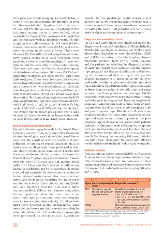
nerve, and tibial nerve recording the motor nerve Table 1: Electro‑physiological types diagnostic standard
conduction velocity, distal latency, and amplitude, Types Electro‑physiological Diagnostic standard %
etc., and observed whether there was a nerve index
conduction block (CB) or not. Sensory conduction AIDP Conduction velocity At least 2 nerves have 1 or more
far‑end lurk waveform electro‑physiological behaviors
test were performed at median nerve, ulnar nerve, disperse F wave < 90 LLN; if CMAP < 50 LLN;
sural nerve, and phil shallow nerve recording the < 85 LLN; > 110 ULN; if CMAP < LLN;
sensory nerve conduction velocity; (2) all patients > 120 ULN; clear waveform disperse
> 120 ULN
had F-wave detection on the median nerve, ulnar AMAN No electro‑physiological behaviors of
nerve peroneal nerve and tibial nerve by recording the AIDP; 2 or more nerves CMAP< 20
wave rate, latency, etc.; (3) needle electromyography AIDP: acute inflammatory demyelinating poly‑neuropathy; AMAN: acute motor
axonal neuropathy; LLN: lower limit of normal; ULN: upper limit of normal;
were performed on thenar muscle, hypothenar CMAP: compound muscle action potential
78 Neuroimmunol Neuroinflammation | Volume 1 | Issue 2 | September 2014