
Page 28 - Read Online
P. 28
Page 8 of 18 Llamoza-Torres et al. Metab Target Organ Damage 2024;4:40 https://dx.doi.org/10.20517/mtod.2024.64
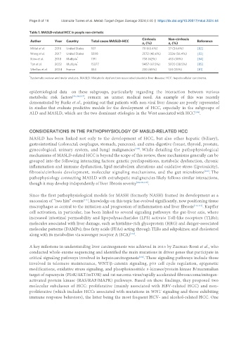
Table 1. MASLD-related HCC in people non-cirrhotic
Cirrhosis Non-cirrhosis
Author Year Country Total cases MASLD-HCC Reference
n, (%) n, (%)
Mittal et al. 2016 United States 107 70 (65.4%) 37 (34.6%) [82]
Wong et al. 2017 United States 5898 2572 (43,6%) 3326 (56.4%) [83]
*
Stine et al. 2018 Multiple 1191 738 (62%) 453 (38%) [84]
Tan et al. 2022 Multiple * 15377 9457 (61.5%) 5920 (38.5%) [85]
Vitellius et al. 2024 France 354 230 (65%) 124 (35%) [81]
*
Systematic review and meta-analysis. MASLD: Metabolic dysfunction-associated steatotic liver disease; HCC: hepatocellular carcinoma.
epidemiological data on these subgroups, particularly regarding the interaction between various
metabolic risk factors [39,106,107] , remain an unmet medical need. An example of this was recently
demonstrated by Burke et al., pointing out that patients with non-viral liver disease are poorly represented
in studies that evaluate predictive models for the development of HCC, especially in the subgroups of
[108]
ALD and MASLD, which are the two dominant etiologies in the West associated with HCC .
CONSIDERATIONS IN THE PATHOPHYSIOLOGY OF MASLD-RELATED HCC
MASLD has been linked not only to the development of HCC, but also other hepatic (biliary),
gastrointestinal (colorectal, esophagus, stomach, pancreas), and extra-digestive (breast, thyroid, prostate,
gynecological, urinary system, and lung) malignancies . While detailing the pathophysiological
[109]
mechanisms of MASLD-related HCC is beyond the scope of this review, these mechanisms generally can be
grouped into the following interacting factors: genetic predispositions, metabolic dysfunction, chronic
inflammation and immune dysfunction, lipid metabolism alterations and oxidative stress (lipotoxicity),
fibrosis/cirrhosis development, molecular signaling mechanisms, and the gut microbiome . The
[109]
pathophysiology connecting MASLD with extrahepatic malignancies likely follows similar interactions,
though it may develop independently of liver fibrosis severity [80,109,110] .
Since the first pathophysiological models for MASH (formerly NASH) framed its development as a
succession of “two hits” events , knowledge on this topic has evolved significantly, now positioning tissue
[111]
macrophages as central to the initiation and progression of inflammation and liver fibrosis [112,113] . Kupffer
cell activation, in particular, has been linked to several signaling pathways: the gut-liver axis, where
increased intestinal permeability and lipopolysaccharides (LPS) activate Toll-like receptors (TLRs);
molecules associated with liver damage, such as histidine-rich glycoprotein (HRG) and danger-associated
molecular patterns (DAMPs); free fatty acids (FFAs) acting through TLRs and adipokines; and cholesterol
along with its metabolites via scavenger receptor A (SCA) .
[112]
A key milestone in understanding liver carcinogenesis was achieved in 2015 by Zucman-Rossi et al., who
conducted whole-exome sequencing and identified the main mutations in driver genes that participate in
[114]
critical signaling pathways involved in hepatocarcinogenesis . These signaling pathways include those
involved in telomere maintenance, WNT/β-catenin signaling, p53 cell cycle regulation, epigenetic
modifications, oxidative stress signaling, and phosphoinositide 3-kinases/protein kinase B/mammalian
target of rapamycin (PI3K/AKT/mTOR) and rat sarcoma virus/rapidly accelerated fibrosarcoma/mitogen-
activated protein kinase (RAS/RAF/MAPK) pathways. Based on these findings, they proposed two
molecular subclasses of HCC: proliferative (mainly associated with HBV-related HCC) and non-
proliferative (which includes HCCs associated with mutations in WNT signaling and those exhibiting
immune response behaviors), the latter being the most frequent HCV- and alcohol-related HCC. One