
Page 340 - Read Online
P. 340
Page 6 of 12 Maalouly et al. Mini-invasive Surg 2021;5:35 https://dx.doi.org/10.20517/2574-1225.2021.57
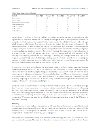
Table 2. Group characteristics of the study
Variable Group A (9) Group B (9) Group C (8) Group D (6) Group E (9)
Screws inserted 56 60 46 38 50
Screws revised 0 1 1 0 3
Intraoperative trajectory revision 0 1 2 1 1
Unreachable trajectory 6 2 0 0 0
Grade A + B Screws 54 58 42 29 43
Grade C screws 2 2 4 9 7
Grade D, E screws 0 0 0 0 0
around Group C. In Group A, the robot could not reach all the planned screw trajectories requiring it to be
repositioned in four cases. The reason was a steep sacral slope in three of these patients which was not
accounted for while planning the trajectory of the screw, causing the robot arm to press on the rib cage,
which obstructed achieving the final trajectory. In one other patient, the robot arm was obstructed by the
operating table frame in the lateral position surgery. The unachieved trajectories were cannulated by hand
using the navigation function of the robot system. No repositioning was needed in the following case groups
as understanding and experience improved the trajectory planning. The surgeon showed a serial decrease in
time taken to plan at each level, with an average of 4.1 min of planning per level. As this was part of the
learning curve of the surgeon with this new technology, all planning and screw placement were done by the
surgeon. Cases in Group E reported a longer planning time of 5.08 min per level due to educating and
teaching of visiting surgeons. The two outlier cases where teaching occurred were omitted, and the
scatterplot obtained showed a decrease in planning time [Figure 3].
In total, 250 screws were inserted using the robotic arm guidance to fix 86 motion segments. Fourteen
screws were inserted without the use of the robot arm. The robot failed to reach its trajectory in eight
screws, and they were inserted using the navigation system of the ExcelsiusGPS robot. Five screws required
an intraoperative adjustment of trajectory. Five screws had to be revised. The revised screws were reported
as one in Group B, on in Group C, and three in Group E. No conclusion could be derived from the
increasing frequency of revised screws in the last group. However, the average insertion time of screws
using the robot showed a decreasing trend [Figure 4].
The mean radiation characteristics of our experience are summarized in Table 3. The mean fluoroscopy
dose in seconds per case was around 16.32 ± 13.22 s with the mean effective dose of 8.2 ± 3.74 mSv during
the 3D CT scan. One patient needed a repeat scan as the CT scan was inverted and could not be validated
by the robot. Fluoroscopy exposure was high in cases that required manual insertion, causing multiple shots
to evaluate screw positioning. Moreover, a high radiation dose was recorded in the three cases where the
preoperative CT protocol was used due to difficulty of fluoroscopy integration to match the CT images to
the patient’s anatomy.
Accuracy
In total, 250 screws were analyzed for accuracy on CT scan. S2 alar iliac screws as part of lumbosacral
construct were deemed acceptable if they did not breach the sacral foramen or breach the pelvic wall. The
overall accuracy in this study was 98%. Overall, 245 screws were deemed acceptable with 226/250 (90.4%)
being Grade A + B and 24/250 (9.6%) being Grade C [Figure 5].
Five screws were revised immediately after confirming on fluoroscopy and before rod placements. Those
were considered inaccurate. The inability of the robot to reach screw trajectory was high in Group A cases